

Colocalization of the IL-12 receptor and FcgammaRIIIa to natural killer cell lipid rafts leads to activation of ERK and enhanced production of interferon-gamma
- PMID: 18174382
- PMCID: PMC2288725
- DOI: 10.1182/blood-2007-01-068908
Colocalization of the IL-12 receptor and FcgammaRIIIa to natural killer cell lipid rafts leads to activation of ERK and enhanced production of interferon-gamma
Abstract
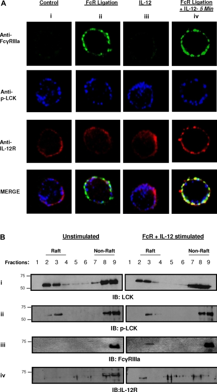
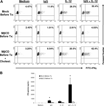
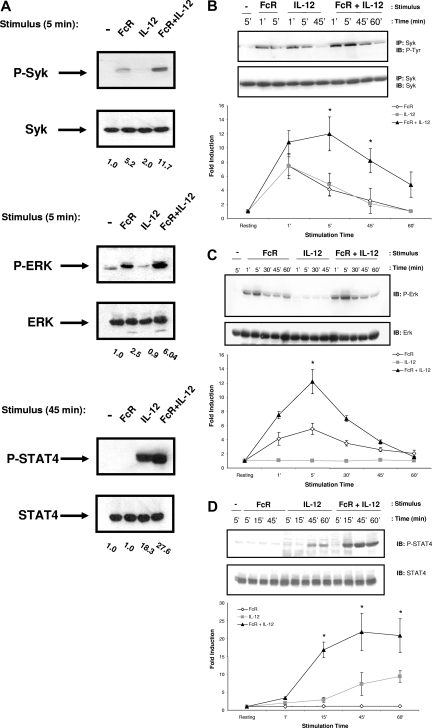
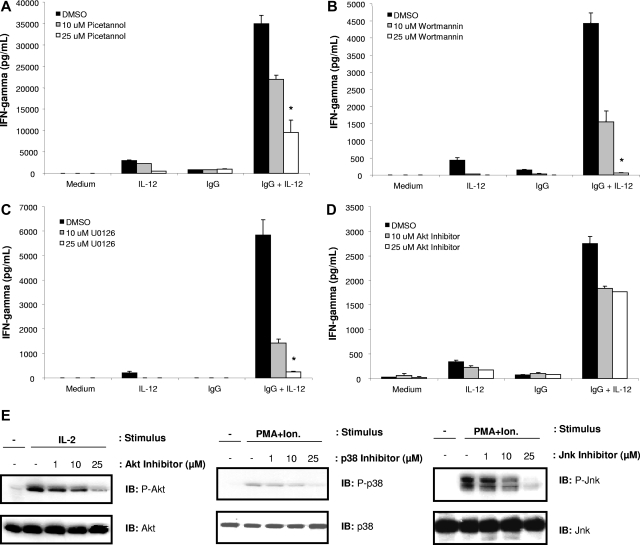
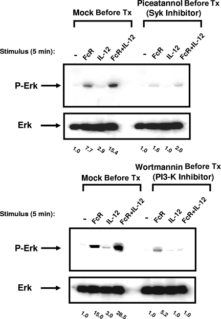
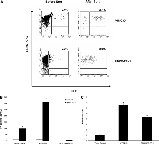
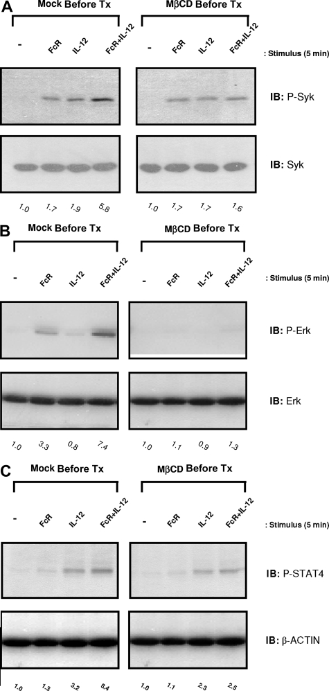
Natural killer (NK) cells express an activating receptor for the Fc portion of IgG (FcgammaRIIIa) that mediates interferon (IFN)-gamma production in response to antibody (Ab)-coated targets. We have previously demonstrated that NK cells activated with interleukin-12 (IL-12) in the presence of immobilized IgG secrete 10-fold or more higher levels of IFN-gamma as compared with stimulation with either agent alone. We examined the intracellular signaling pathways responsible for this synergistic IFN-gamma production. NK cells costimulated via the FcR and the IL-12 receptor (IL-12R) exhibited enhanced levels of activated STAT4 and Syk as compared with NK cells stimulated through either receptor alone. Extracellular signal-regulated kinase (ERK) was also synergistically activated under these conditions. Studies with specific chemical inhibitors revealed that the activation of ERK was dependent on the activation of PI3-K, whose activation was dependent on Syk, and that sequential activation of these molecules was required for NK cell IFN-gamma production in response to FcR and IL-12 stimulation. Retroviral transfection of ERK1 into primary human NK cells substantially increased IFN-gamma production in response to immobilized IgG and IL-12, while transfection of human NK cells with a dominant-negative ERK1 abrogated IFN-gamma production. Confocal microscopy and cellular fractionation experiments revealed that FcgammaRIIIa and the IL-12R colocalized to areas of lipid raft microdomains in response to costimulation with IgG and IL-12. Chemical disruption of lipid rafts inhibited ERK signaling in response to costimulation and significantly inhibited IFN-gamma production. These data suggest that dual recruitment of FcgammaRIIIa and the IL-12R to lipid raft microdomains allows for enhanced activation of downstream signaling events that lead to IFN-gamma production.
Figures
Comment in
-
Rafting with the IL-12 receptor.Blood. 2008 Apr 15;111(8):3911-2. doi: 10.1182/blood-2008-01-134627. Blood. 2008. PMID: 18434959 Free PMC article.
Similar articles
-
Src homology 2-containing inositol 5'-phosphatase 1 negatively regulates IFN-gamma production by natural killer cells stimulated with antibody-coated tumor cells and interleukin-12.Cancer Res. 2005 Oct 1;65(19):9099-107. doi: 10.1158/0008-5472.CAN-04-4424. Cancer Res. 2005. PMID: 16204085
-
CD160-activating NK cell effector functions depend on the phosphatidylinositol 3-kinase recruitment.Int Immunol. 2007 Apr;19(4):401-9. doi: 10.1093/intimm/dxm005. Epub 2007 Feb 16. Int Immunol. 2007. PMID: 17307798
-
Effects of interleukin-18 on natural killer cells: costimulation of activation through Fc receptors for immunoglobulin.Cancer Immunol Immunother. 2013 Jun;62(6):1073-82. doi: 10.1007/s00262-013-1403-0. Epub 2013 Apr 19. Cancer Immunol Immunother. 2013. PMID: 23604103 Free PMC article.
-
Impact of Plasma Membrane Domains on IgG Fc Receptor Function.Front Immunol. 2020 Jun 30;11:1320. doi: 10.3389/fimmu.2020.01320. eCollection 2020. Front Immunol. 2020. PMID: 32714325 Free PMC article. Review.
-
Immunology of IL-12: An update on functional activities and implications for disease.EXCLI J. 2020 Dec 11;19:1563-1589. doi: 10.17179/excli2020-3104. eCollection 2020. EXCLI J. 2020. PMID: 33408595 Free PMC article. Review.
Cited by
-
miR-155 regulates IFN-γ production in natural killer cells.Blood. 2012 Apr 12;119(15):3478-85. doi: 10.1182/blood-2011-12-398099. Epub 2012 Feb 29. Blood. 2012. PMID: 22378844 Free PMC article.
-
Interleukin (IL)-23 Stimulates IFN-γ Secretion by CD56bright Natural Killer Cells and Enhances IL-18-Driven Dendritic Cells Activation.Front Immunol. 2018 Jan 17;8:1959. doi: 10.3389/fimmu.2017.01959. eCollection 2017. Front Immunol. 2018. PMID: 29403472 Free PMC article.
-
IL-21 Enhances Natural Killer Cell Response to Cetuximab-Coated Pancreatic Tumor Cells.Clin Cancer Res. 2017 Jan 15;23(2):489-502. doi: 10.1158/1078-0432.CCR-16-0004. Epub 2016 Jul 19. Clin Cancer Res. 2017. PMID: 27435400 Free PMC article.
-
A peptide inhibitor of antibody-dependent cell-mediated cytotoxicity against EGFR/folate receptor-α double positive cells.Medchemcomm. 2018 Feb 26;9(5):783-788. doi: 10.1039/c8md00010g. eCollection 2018 May 1. Medchemcomm. 2018. PMID: 30108967 Free PMC article.
-
Co-stimulation of the fc receptor and interleukin-12 receptor on human natural killer cells leads to increased expression of cd25.Oncoimmunology. 2017 Oct 16;7(2):e1381813. doi: 10.1080/2162402X.2017.1381813. eCollection 2018. Oncoimmunology. 2017. PMID: 29308301 Free PMC article.
References
-
- Young HA, Ortaldo J. Cytokines as critical co-stimulatory molecules in modulating the immune response of natural killer cells. Cell Res. 2006;16:20–24. - PubMed
-
- Parihar R, Nadella P, Lewis A, et al. A phase I study of interleukin 12 with trastuzumab in patients with human epidermal growth factor receptor-2-overexpressing malignancies: analysis of sustained interferon gamma production in a subset of patients. Clin Cancer Res. 2004;10:5027–5037. - PubMed
-
- Carson WE, Roda J, Parihar R, Lamb T, Shapiro C, Bekaii-Saab T. Phase I trial of interleukin-12 with trastuzumab and paclitaxel in HER2-overexpressing malignancies. J Clin Oncol. 2005;23:173s.
-
- National Center for Biotechnology Information. [Accessed September 6, 2006]. GenBank [database] Available: http://www.ncbi.nlm.nih.gov/Genbank.
Publication types
MeSH terms
Substances
Grants and funding
LinkOut - more resources
Full Text Sources
Other Literature Sources
Miscellaneous
