. 2008 Feb 1;319(5863):630-3.
doi: 10.1126/science.1151298. Epub 2008 Jan 3.

Direct observation of hierarchical folding in single riboswitch aptamers

Affiliations

Direct observation of hierarchical folding in single riboswitch aptamers

William J Greenleaf et al. Science. .

Abstract

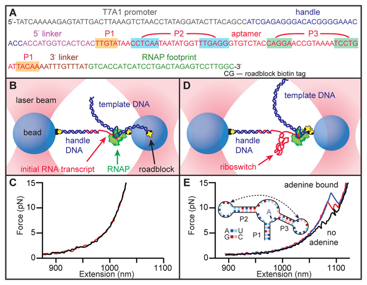
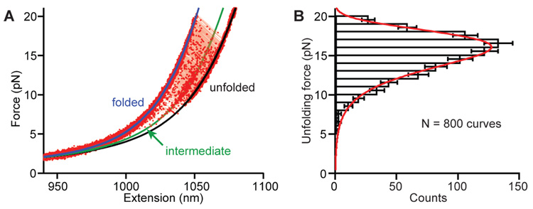
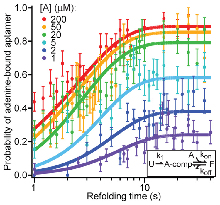
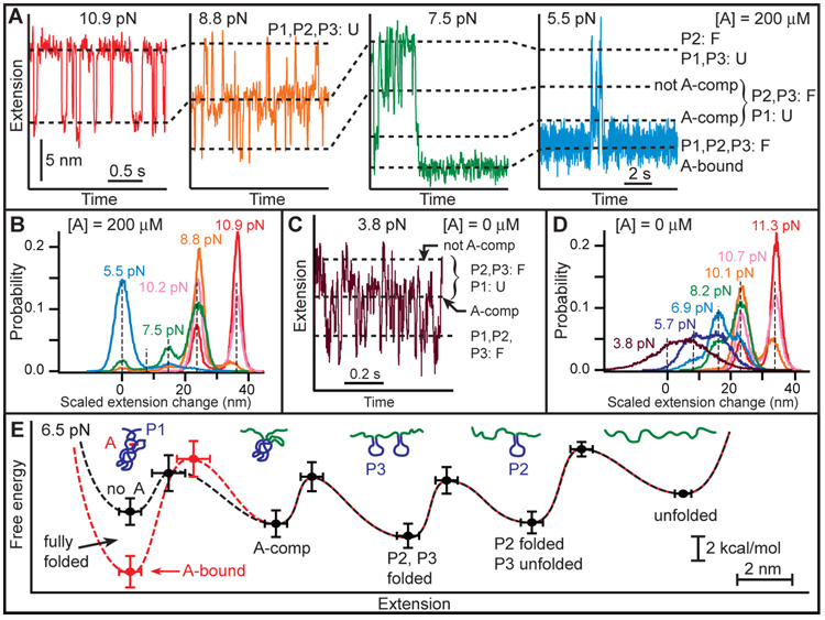
Riboswitches regulate genes through structural changes in ligand-binding RNA aptamers. With the use of an optical-trapping assay based on in situ transcription by a molecule of RNA polymerase, single nascent RNAs containing pbuE adenine riboswitch aptamers were unfolded and refolded. Multiple folding states were characterized by means of both force-extension curves and folding trajectories under constant force by measuring the molecular contour length, kinetics, and energetics with and without adenine. Distinct folding steps correlated with the formation of key secondary or tertiary structures and with ligand binding. Adenine-induced stabilization of the weakest helix in the aptamer, the mechanical switch underlying regulatory action, was observed directly. These results provide an integrated view of hierarchical folding in an aptamer, demonstrating how complex folding can be resolved into constituent parts, and supply further insights into tertiary structure formation.


Figures

Fig. 1
Fig. 1
(A) DNA template used for RNA transcription, showing the sequence of the non-transcribed promoter, the 25-bp section hybridizing with the DNA handle, the pbuE riboswitch aptamer (base-paired helices highlighted) flanked by short linkers, and the footprint of RNAP when stalled by the terminal roadblock. (B) Schematic of the optical trapping assay showing experimental geometry, with stalled RNAP and initial RNA transcript hybridized to the dsDNA handle (not to scale). (C) Two FECs obtained prior to aptamer transcription show little or no structure in the initial transcript. (D) Template DNA is transcribed in situ, producing an aptamer transcript, after which RNAP is stalled by a streptavidin molecule bound to the biotin-based roadblock. (E) FECs obtained after transcription show unfolding transitions in the aptamer. Without adenine, two events are seen (black), corresponding to the unfolding of hairpins P2 and P3 (inset). With adenine bound to the aptamer, larger unfolding events are observed (blue), sometimes involving an intermediate state (red).
Fig. 2
Fig. 2
(A) Non-equilibrium FECs for folded aptamer display a wide distribution of unfolding forces. WLC fit to the folded state (blue), and double WLC fits to the intermediate (green) and unfolded (black) states, indicate contour length changes of 39 ± 1 nt and 62 ± 1 nt for unfolding to the intermediate and unfolded states, respectively. (B) The unfolding force distribution is fit by a model returning the unfolding rate, along with the location and height of the energy barrier to unfolding.
Fig. 3
Fig. 3
Kinetics of aptamer refolding and binding. The fraction of FECs corresponding to the fully-folded, adenine-bound aptamer (identified by the appropriate unfolding signature) for various adenine concentrations as a function of the variable time delay for refolding between pulls. Solid curves display the global fit to a minimal 3-state kinetic scheme (inset): “U” = unfolded, “A-comp” = competent to bind adenine, “F” = folded; adenine-bound.
Fig. 4
Fig. 4
Aptamer states and energetics determined by refolding at constant force. (A) As force is reduced, first P2 refolds (red), then P3 folds (orange). At lower forces, P2 and P3 interact to form a binding pocket and adenine binds, generating two additional states (green). The adenine-bound state is stable over many seconds, even at 5 pN load (blue). (B) Histograms of complete trajectories at different forces, with extension changes scaled by the force-dependent extension per nucleotide. Dashed lines indicate distinct states; the A-comp state is rarely populated. (C, D) Refolding trajectory and histograms in the absence of adenine. P2 and P3 folding occur as with adenine, but the A-comp state is now highly populated at low force, while the folded state is very unstable, even at low force (purple). (E) Quantitative energy landscapes for aptamer folding at 6.5 pN, reconstructed from the experimental data in the presence (red) and absence (black) of adenine. The five potential wells correspond to five observed folding states, illustrated by cartoons. Adenine binding only significantly affects the barrier and energy of the folded state.

References

    1. Winkler WC, Breaker RR. Annu. Rev. Microbiol. 2005;59:487. - PubMed
    1. Coppins RL, Hall KB, Groisman EA. Curr. Opin.Microbiol. 2007;10:176. - PMC - PubMed
    1. Batey RT, Gilbert SD, Montange RK. Nature. 2004;432:411. - PubMed
    1. Serganov A, et al. Chem. Biol. 2004;11:1729. - PMC - PubMed
    1. Mandalb M, Boese B, Barrick JE, Winkler WC, Breaker RR. Cell. 2003;113:577. - PubMed

Publication types