

Intravitreal triamcinolone acetonide inhibits breakdown of the blood-retinal barrier through differential regulation of VEGF-A and its receptors in early diabetic rat retinas
- PMID: 18174522
- PMCID: PMC2836241
- DOI: 10.2337/db07-0982
Intravitreal triamcinolone acetonide inhibits breakdown of the blood-retinal barrier through differential regulation of VEGF-A and its receptors in early diabetic rat retinas
Erratum in
- Diabetes. 2010 Mar;59(3):756
Abstract
Objective: To elucidate the mechanism of the unique beneficial effect of intravitreal steroid therapy on diabetic macular edema, we investigated the effect of locally administered triamcinolone acetonide (TA) on the expression of vascular endothelial growth factor (VEGF)-A and its receptors in retinas of rats with streptozotocin (STZ)-induced diabetes. We then correlated the expression of these proteins with breakdown of the blood-retinal barrier (BRB).
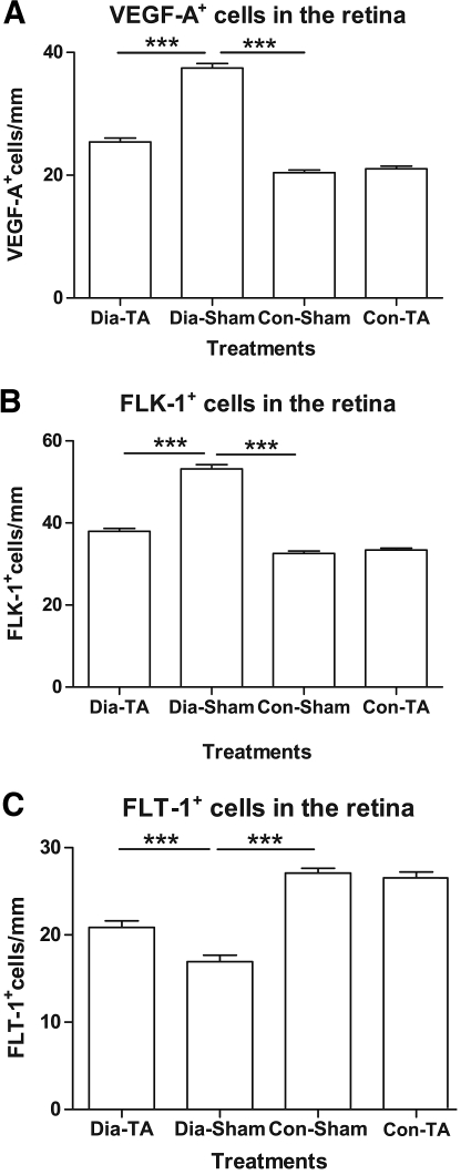
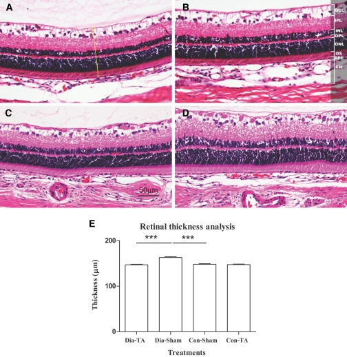
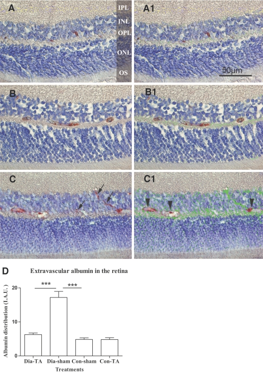
Research design and methods: Thirty-two eyes of 16 diabetic and nondiabetic rats were divided into four groups. TA was injected into the vitreous of the right eye, and saline was injected into the left eye (control) 3.5 weeks after induction of diabetes. Retinas were harvested 48 h following treatment. mRNA and protein expression of VEGF-A, VEGF-A receptor 1 (fms-like tyrosine kinase [FLT]-1), and VEGF-A receptor 2 (fetal liver kinase [FLK]-1) were determined by real-time RT-PCR and immunohistochemistry. BRB permeability was quantitated by measuring extravasated endogenous albumin and retinal thickness.
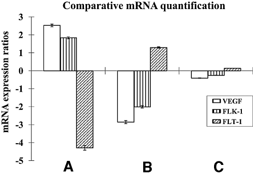
Results: Diabetes-induced retinal thickness and albumin extravasation were significantly reduced in TA-treated diabetic retinas to a level similar to that in sham-treated nondiabetic eyes. A close correlation between albumin leakage and increased expression of both Vegf-a and Flk-1 was noted in the diabetic retinas. TA downregulated the expression of Vegf-a and Flk-1 but upregulated the expression of Flt-1. TA did not alter the expression of these genes in nondiabetic retinas.
Conclusions: Intravitreal injection of TA stabilizes the BRB in association with regulation of Vegf-a, Flk-1, and Flt-1 expression in retinas in the early stages of diabetes.
Figures
References
-
- Kohner EM: Diabetic retinopathy and high blood pressure: defining the risk. Am J Hypertens 10:181S–183S, 1997 - PubMed
-
- Group ETDRSR: Effects of asprin treatment on diabetic retinopathy ETDRS report number 8. Opthalmology 98 (Suppl.):757–765, 1991 - PubMed
-
- Sander B, Larsen M, Moldow B, Lund-Andersen H: Diabetic macular edema: passive and active transport of fluorescein through the blood-retina barrier. Invest Ophthalmol Vis Sci 42:433–438, 2001 - PubMed
-
- Engler CB, Sander B, Larsen M, Koefoed P, Parving HH, Lund-Andersen H: Probenecid inhibition of the outward transport of fluorescein across the human blood-retina barrier. Acta Ophthalmol (Copenh) 72:663–667, 1994 - PubMed
-
- Vinores SA, McGehee R, Lee A, Gadegbeku C, Campochiaro PA: Ultrastructural localization of blood-retinal barrier breakdown in diabetic and galactosemic rats. J Histochem Cytochem 38:1341–1352, 1990 - PubMed
Publication types
MeSH terms
Substances
LinkOut - more resources
Full Text Sources
Medical
Miscellaneous
