Review
. 2007 Nov 30:5:e011.
doi: 10.1621/nrs.05011.

Steroid receptor coactivator 2 is required for female fertility and mammary morphogenesis: insights from the mouse, relevance to the human

Affiliations
Review

Steroid receptor coactivator 2 is required for female fertility and mammary morphogenesis: insights from the mouse, relevance to the human

Atish Mukherjee et al. Nucl Recept Signal. .

Abstract

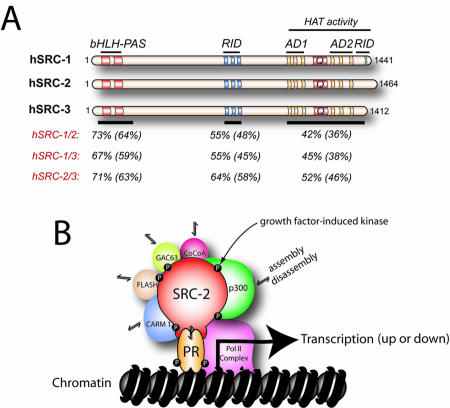
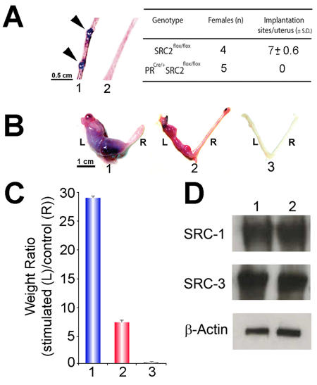
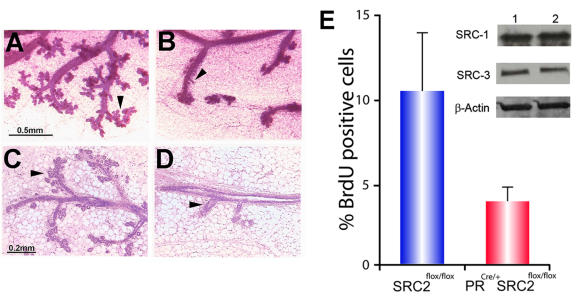
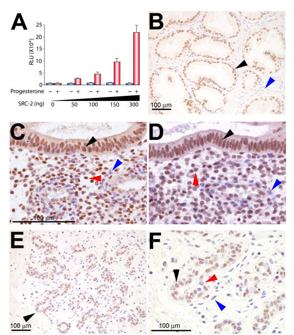
Although the importance of the progesterone receptor (PR) to female reproductive and mammary gland biology is firmly established, the coregulators selectively co-opted by PR in these systems have not been clearly delineated. A selective gene-knockout approach applied to the mouse, which abrogates gene function only in cell types that express PR, recently disclosed steroid receptor coactivator 2 (SRC-2, also known as TIF-2 or GRIP-1) to be an indispensable coregulator for uterine and mammary gland responses that require progesterone. Uterine cells positive for PR (but devoid of SRC-2) were found to be incapable of facilitating embryo implantation, a necessary first step toward the establishment of the materno-fetal interface. Importantly, such an implantation defect is not exhibited by knockouts for SRC-1 or SRC-3, underscoring the unique coregulator importance of SRC-2 in peri-implantation biology. Moreover, despite normal levels of PR, SRC-1 and SRC-3, progesterone-dependent branching morphogenesis and alveologenesis fails to occur in the murine mammary gland in the absence of SRC-2, thereby establishing a critical coregulator role for SRC-2 in signaling cascades that mediate progesterone-induced mammary epithelial proliferation. Finally, the recent detection of SRC-2 in the human endometrium and breast suggests that this coregulator may represent a new clinical target for the future management of female reproductive health and/or breast cancer.


Figures

Figure 1
Figure 1. The SRC/p160 family of coregulators.
Panel A shows the domain structure of human (h) SRC-1, -2, and -3 proteins. The basic helix loop helix, Per/ARNT/Sim, receptor interaction and activation domains are indicated by bHLH, PAS, RID, and AD, respectively. The amino acid region associated with histone acetyl transferase (HAT) activity in SRC-1 and -3 is also denoted; Q indicates the glutamine-rich region. Also shown is the similarity and (identity) of amino acid sequences within key functional domains of SRC members. Overall amino acid similarity and (identity) between SRC members is: hSRC1/2, 54% (46%); hSRC1/3, 50% (43%); and hSRC2/3, 55% (48%). Amino acid sequence alignments were conducted using LALNVIEW software (Duret et al., 1996). Panel B shows an schematic model in which the pairing of SRC-2 with PR at the genome comprises part of a dynamic multiprotein transcriptional complex in which such co-coregulators as p300 (Chen et al., 2000a), CARM-1 (Chen et al., 1999), FLASH (Kino et al., 2004), GAC63 (Chen et al., 2005), and CoCoA (Kim and Stallcup, 2004) differentially assemble and disassemble depending on a particular input signal, such as a distinct phosphorylation event mediated by a growth factor or cell survival signal-induced kinase.
Figure 2
Figure 2. Abrogation of uterine SRC-2 results in a block in embryo implantation and a partial decidual response.
In panel A, arrows show the location of implantation sites in the uterus (1) of a SRC-2flox/flox (or wild-type (WT)) mouse (5.5 days post coitum (d.p.c.)). However, implantation sites were not detected in uteri from similarly treated PRCre/+SRC-2flox/flox (2) mice. The average number of implantation sites per genotype per total number of mice examined is tabulated. In panel B, the gross morphological response of the left (L) uterine horn to a deciduogenic stimulus for SRC-2flox/flox (1), PRCre/+SRC-2flox/flox (2), and PRCre/+SRC-2flox/flox SRC-1KO trigenic (3) mice is shown. The right (R) uterine horn represents the unstimulated control. Although the PRCre/+SRC-2flox/flox uterus (2) exhibits a limited decidual response, note the absence of a decidual response in the PRCre/+SRC-2flox/flox SRC-1KO trigenic uterus (3). Panel C graphically presents the average weight ratios (± standard deviation (SD)) of stimulated (L) to control (R) horn for SRC-2flox/flox (1), PRCre/+SRC-2flox/flox (2), and PRCre/+SRC-2flox/flox SRC-1KO trigenic (3) uteri. Western analysis in panel D reveals uterine tissue from untreated adult virgin SRC-2flox/flox (1) and PRCre/+SRC-2flox/flox (2) mice show equivalent levels of uterine SRC-1 and SRC-3 (loading control is β-actin). Modified from (Mukherjee et al., 2006b) (Copyright (2006) American Society for Microbiology).
Figure 3
Figure 3. Absence of mammary SRC-2 function blocks progestin-induced ductal side-branching and alveologenesis.
Panels A and B show whole-mounts of mammary glands from SRC-2flox/flox and PRCre/+SRC-2flox/flox mice (following three weeks of estrogen plus progesterone (EP) exposure), respectively. Unlike the SRC-2flox/flox mammary gland (positive control), note the marked reduction in branching morphogenesis (black arrow) in the PRCre/+SRC-2flox/flox gland. Panels C and D represent hematoxylin and eosin (H&E) stained sections of tissue shown in panels A and B, respectively. Compared to the SRC-2flox/flox gland (panel C), note the conspicuous reduction in the epithelial compartment in the PRCre/+SRC-2flox/flox gland (panel D (arrowhead)). The graph in panel E displays the average percentage of mammary epithelial cells (± standard deviation (S.D.)) positive for BrdU incorporation in the hormone-treated SRC-2flox/flox and PRCre/+SRC-2flox/flox glands. Inset shows an SRC-2flox/flox (1) and PRCre/+SRC-2flox/flox (2) immunoblot for mammary SRC-1 and -3. In comparison to SRC-2flox/flox, changes in SRC-1 and -3 protein levels are not observed in the PRCre/+SRC-2flox/flox mammary gland (β-actin acts as a loading control). Scale bars in panels A and C apply to B and D, respectively. Modified from (Mukherjee et al., 2006b) (Copyright (2006) American Society for Microbiology).
Figure 4
Figure 4. Steroid hormone responsive tissues express SRC-2 in the human.
Panel A shows the increase in ligand-dependent transactivational potency of human PR-B is dependent on increased levels of human SRC-2 (red bars ± S.D.); in the absence of ligand, this increase is not observed (blue bars). For these experiments, human PR-B; SRC-2 (both cloned into pCR3.1) and the luciferase reporter pGRE.E1b.LUC were transiently cotransfected into HeLa cells in the presence or absence of 10-7M R5020, as described previously (Lonard et al., 2004); results are expressed in relative light units (RLU). Panel B shows SRC-2 is expressed in the majority of epithelial cells of the human prostate (black arrow), an established cellular target for androgen receptor action (Culig et al., 2002); note: the stromal compartment registers negative for SRC-2 expression (blue arrow). Panels C and D show transverse sections of the luminal and stromal compartment (with surrounding stroma) of the human endometrium stained for PR and SRC-2 expression, respectively. Note that PR and SRC-2 are detected in nuclei of the same cell types in both cellular compartments (black and red arrows, respectively). The blue arrow in panels C and D highlights a stromal cell negative for PR and SRC-2 expression, respectively; scale bar in panel C applies to panel D. Endometrial biopsies were obtained by endometrial pipelle from healthy women with normal cycles (aged between 18-35 years) during the mid-secretory (luteal) phase of the menstrual cycle (days 20-24, which is based on the ideal 28 day cycle, in which day 1 represents the first day of menstrual flow and day 14 the day of ovulation); cycle phase was determined relative to the timing of the urinary luteinizing hormone (LH) surge. Immunohistochemical detection of human SRC-2 and PR was undertaken using established methods previously reported by our group (Lee et al., 2005; Mukherjee et al., 2006b). Panel E shows a representative example of a normal type 1 terminal ductal lobular unit (TDLU) of the human breast in which SRC-2 expression is restricted to the epithelial compartment (black arrow). Panel F is a higher magnification of the region indicated by the black arrow in panel E. Note that SRC-2 expression is confined to a subset of epithelial cells of the TDLU (black arrow indicates an epithelial cell scoring positive for SRC-2 expression, whereas the red arrow highlights an epithelial cell which is negative for SRC-2 expression; blue arrow denotes a stromal cell which is negative for SRC-2 expression). Interestingly, the spatial expression pattern of mammary SRC-2 resembles that previously reported for ER-α and PR in the human breast (Clarke et al., 1997). With institutional review board approval, human tissue samples were obtained from Baylor College of Medicine affiliated hospitals. Modified from (Mukherjee et al., 2006a) (Copyright (2006) Elsevier, B.V.).

Similar articles

Cited by

References

    1. Anzick S. L., Kononen J., Walker R. L., Azorsa D. O., Tanner M. M., Guan X. Y., Sauter G., Kallioniemi O. P., Trent J. M., Meltzer P. S. AIB1, a steroid receptor coactivator amplified in breast and ovarian cancer. Science. 1997;277:965–8. - PubMed
    1. Belandia B., Parker M. G. Functional interaction between the p160 coactivator proteins and the transcriptional enhancer factor family of transcription factors. J Biol Chem. 2000;275:30801–5. - PubMed
    1. Berrevoets C. A., Umar A., Trapman J., Brinkmann A. O. Differential modulation of androgen receptor transcriptional activity by the nuclear receptor co-repressor (N-CoR) Biochem J. 2004;379:731–8. - PMC - PubMed
    1. Chen Y. H., Kim J. H., Stallcup M. R. GAC63, a GRIP1-dependent nuclear receptor coactivator. Mol Cell Biol. 2005;25:5965–72. - PMC - PubMed
    1. Chen S. L., Dowhan D. H., Hosking B. M., Muscat G. E. The steroid receptor coactivator, GRIP-1, is necessary for MEF-2C-dependent gene expression and skeletal muscle differentiation. Genes Dev. 2000b;14:1209–28. - PMC - PubMed

Publication types

Substances