Comparative Study
. 2007 Dec;115 Suppl 1(Suppl 1):62-8.
doi: 10.1289/ehp.9367.

Effects of chronic genistein treatment in mammary gland, uterus, and vagina

Affiliations
Comparative Study

Effects of chronic genistein treatment in mammary gland, uterus, and vagina

Guillermo Rimoldi et al. Environ Health Perspect. 2007 Dec.

Abstract

Background: The isoflavone genistein (GEN) is found in soy (Glycine max) and red clover (Trifolium pratense). The estrogenic activity of GEN is known, and it is widely advertised as a phytoestrogen useful in alleviating climacteric complaints and other postmenopausal disorders. Knowledge of effects of long-term administration of GEN in laboratory animals is scarce, and effects in the uterus and mammary gland after long-term administration have not been studied. The uterus and mammary gland are known to be negatively influenced by estrogens used in hormone therapy.

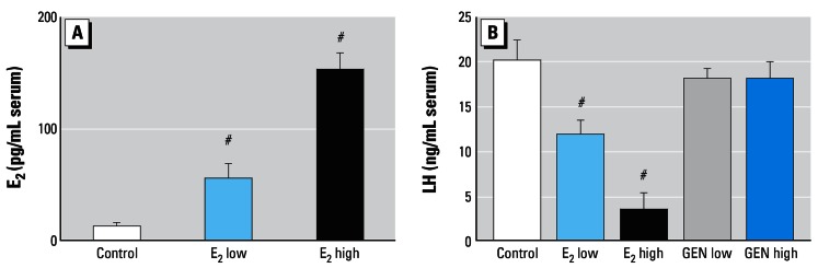
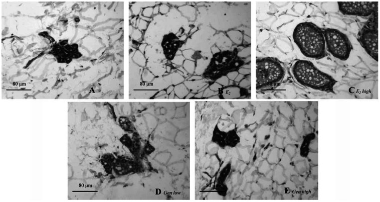
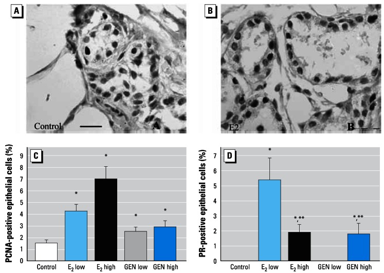
Objectives: We administered two doses of GEN [mean daily uptake 5.4 (low) or 54 mg/kg (high) body weight (bw)] orally over a period of 3 months to ovariectomized (ovx) rats and compared the effects with a treatment with two doses of 17beta-estradiol [E(2); 0.17 (low) or 0.7 mg/kg bw (high)]. Mammary glands, vaginae, and uteri were investigated morphologically and immunohistochemically. We quantified the expression of proliferating cell nuclear antigen (PCNA) and progesterone receptor (PR) in the mammary gland.

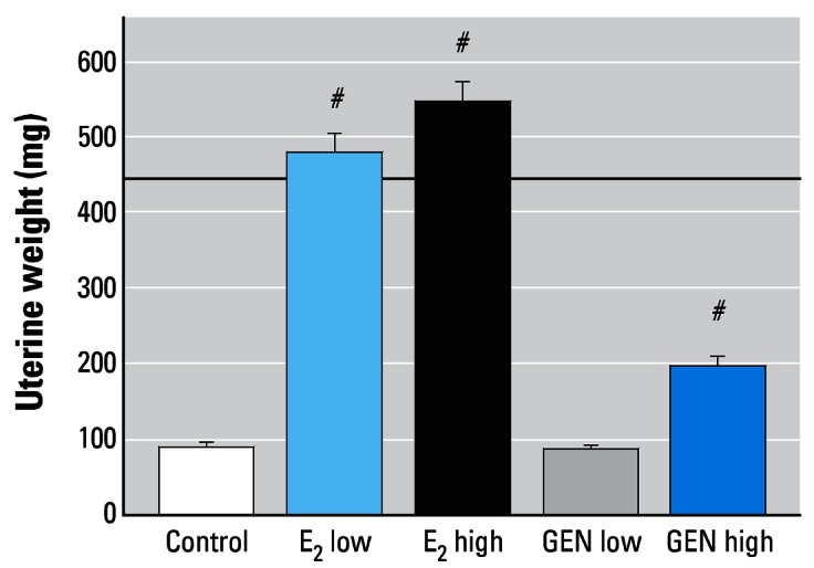
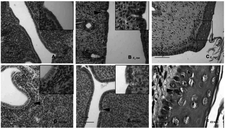
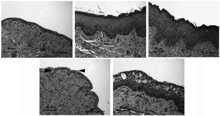
Results: In rats treated with either of the E(2) doses or the high GEN dose, we found increased uterine weight, and histologic analysis showed estrogen-induced features in the uteri. In vaginae, either E(2) dose or GEN high induced hyperplastic epithelium compared with the atrophic controls. In the mammary gland, E(2) (either dose) or GEN increased proliferation and PR expression. Serum levels of luteinizing hormone were decreased by E(2) (both doses) but not by GEN.

Conclusions: In summary, E(2) and GEN share many effects in the studied organs, particularly in the vagina, uterus, and mammary gland but not in the hypothalamo/pituitary unit.

Keywords: estradiol; genistein; mammary gland; uterus; vagina.


Figures

Figure 1
Figure 1
Serum levels (mean ± SEM) of E2 (A) and LH (B) in ovx rats after 3 months of treatment. Note the dose-dependent reduction of LH by E2 and the lacking effect of GEN. #p > 0.01 compared with control.
Figure 2
Figure 2
Uterine wet weights (mean ± SEM) of ovx rats after 3 months of treatment with E2 or GEN. Note the stimulating effects of both E2 doses and the GEN high. The dashed line indicates the average uterine wet weight for an intact female rat. #p < 0.01 compared with control.
Figure 3
Figure 3
Photomicrographs of representative uteri from one animal per treatment group. (A) Control: atrophic uterus with cuboidal endometrial epithelium and loose connective tissue composed of round nuclei in an unorganized pattern. (B) E2 low: the endometrium consists of tall single-layered epithelial cells with abundant mitotic figures and necrosis (arrowhead); stromal cells of endometrial lamina propria are well organized and spindle shaped. (C) E2 high: all structures are hypertrophic and hyperplastic; the endometrial epithelium is multilayered with squamous metaplasia and atypic mitotic figure (arrowhead) surrounded by anaplastic epithelial nuclei, and keratinized cells are present in the lumen. The square in (C) indicates the area shown at higher magnification in (F). (D) GEN low: lamina propria cells adopted an organized pattern (arrowhead), with more condensed nuclei of spindle shape. (E) GEN high: areas of high endometrial proliferation and unclear influences on lamina propria cells are present. Arrowheads indicate cells undergoing mitosis. Insets in (A), (B), (D), and (E) show higher magnifications of endometrium. Bar = 80 μm in (A–E) and 20 μm in (F).
Figure 4
Figure 4
Photomicrographs of representative vaginal sections from one animal per treatment group. (A) Control: atrophic vaginal epithelium is composed of a few layers of flattened cells. (B) E2 low: vaginal epithelium is hypertrophic and hyperplastic, with cornification in the upper layers. (C) E2 high: stronger hypertrophy and hyperplasia of epithelium are present; compared with E2 low, there are more cell layers and a higher degree of cornification. (D) GEN low: atrophic vaginal epithelium and the thickness and number of layers resemble that of control animals, but some areas have an incipient cytoplasmatic vacuolization (arrowheads). (E) GEN high: vaginal epithelium is increased (5–10 cells per layer on average), and large cytoplasmatic vacuoles are present in the upper layers. Bar = 80 μm.
Figure 5
Figure 5
Photomicrographs of representative mammary gland sections from one animal per treatment group. (A) Control: a small cluster of densely packed epithelial cells without luminal formation are present in the deep subcutaneous fat pad. (B) E2 low: a few terminal structures with small lumina but without secretory material. (C) E2 high: well-formed acinar and luminal structures with secretory material in the lumina. In GEN low (D) and GEN high (E), there is almost no cellular or acinar difference from control. Bars = 80 μm.
Figure 6
Figure 6
Results of immunostaining for PCNA (A,C) and PR (B,D) in mammary gland. (A) Photomicrograph showing PCNA immunostaining in control tissue. (B) Photomicrograph showing PR immunostaining in E2 high mammary gland. Percentage of PCNA-positive (C) and PR-expressing (D) epithelial cells in mammary terminal structures. Immunostained PCNA cells were regularly seen in all animals (C). PR-expressing cells were seen in both in E2 groups and in the GEN high group (D); note the reduced number of PR-expressing cells in E2 high versus E2 low mammary glands. Bars = 20 μm. *p < 0.05 compared with control; **p < 0.05 compared with E2 low.

Similar articles

Cited by

References

    1. Albertazzi P, Sharma S. Urogenital effects of selective estrogen receptor modulators: a systematic review. Climacteric. 2005;8(3):214–220. - PubMed
    1. Allred CD, Allred KF, Ju YH, Clausen LM, Doerge DR, Schantz SL, et al. Dietary genistein results in larger MNU-induced, estrogen-dependent mammary tumors following ovariectomy of Sprague-Dawley rats. Carcinogenesis. 2004;25(2):211–218. - PubMed
    1. Asarian L, Geary N. Modulation of appetite by gonadal steroid hormones. Philos Trans R Soc Lond B Biol Sci. 2006;361(1471):1251–1263. - PMC - PubMed
    1. Beral V. Breast cancer and hormone-replacement therapy in the Million Women Study. Lancet. 2003;362(9382):419–427. - PubMed
    1. Boue SM, Wiese TE, Nehls S, Burow ME, Elliott S, Carter-Wientjes CH, et al. Evaluation of the estrogenic effects of legume extracts containing phytoestrogens. J Agric Food Chem. 2003;51(8):2193–2199. - PubMed

Publication types

MeSH terms