. 2008 May;35(5):958-65.
doi: 10.1007/s00259-007-0660-6. Epub 2008 Jan 4.

The diagnostic value of 124I-PET in patients with differentiated thyroid cancer

Affiliations

The diagnostic value of 124I-PET in patients with differentiated thyroid cancer

Ha T T Phan et al. Eur J Nucl Med Mol Imaging. 2008 May.

Abstract

Background: The purpose of this prospective study was to evaluate the clinical diagnostic value of iodine-124 (124I)-positron emission tomography (PET) in patients with advanced differentiated thyroid carcinoma (DTC) and to compare the 124I-PET imaging results with the 131I whole-body scan (WBS).

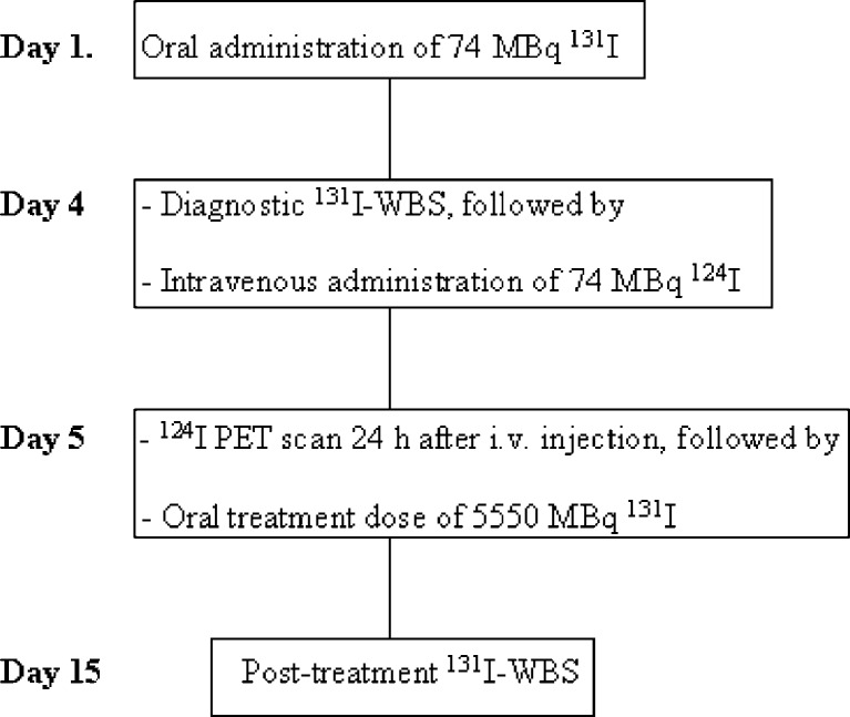
Materials and methods: Twenty patients with histologically proven advanced DTC (including T4, extra-nodal tumour growth, or distant metastases) underwent diagnostic 131I-WBS, 124I-PET scan, and post-treatment 131I-WBS 4 months after ablation. The findings on the 124I-PET were compared with the findings on the diagnostic and post-therapeutic 131I-WBS and were also correlated with radiologic and/or cytological investigations.

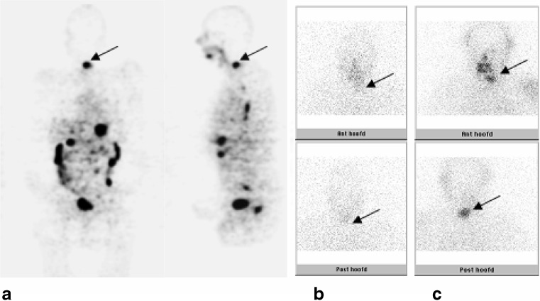
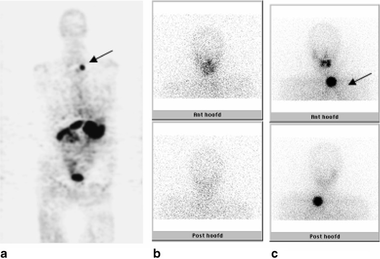
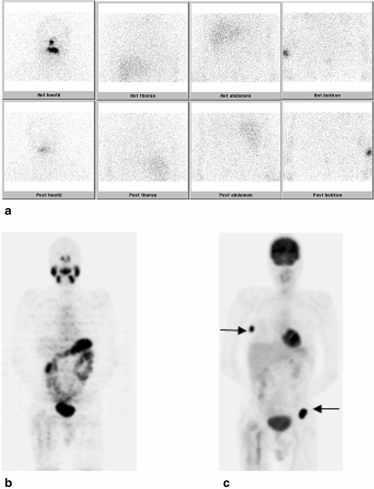
Results: 124 I-PET vs diagnostic 131 I-WBS. Eleven patients showed uptake on the 124I-PET. Only 3 of these 11 patients also showed uptake on the diagnostic 131I scan, but the uptake was more clearly visible and the abnormalities were more extensive on the 124I-PET. 124 I-PET vs post-treatment 131 I-WBS. Eleven patients showed uptake on the 124I-PET, which was also visible on the post-treatment scan in nine patients; in the other two patients, no uptake was observed on the post-treatment scan and no anatomical localisation could be confirmed. Two patients showed only uptake on the post-treatment scan without uptake on the 124I-PET: in one, the uptake was confirmed by MRI, and in the other, no anatomical localisation was found. In seven patients, no uptake was observed on both the scans.

Conclusion: 124I-PET proved to be a superior diagnostic tool as compared to low-dose diagnostic 131I scans and adequately predicted findings on subsequent high-dose post-treatment 131I scans.


Figures

Fig. 1
Fig. 1
The diagram showed a schematic course of the investigations performed 4 months after ablation
Fig. 2
Fig. 2
This patient (no. 1) showed a clearly visible lesion in the cervical vertebrae on the 124I-PET (a, arrow), comparable with the lesion visible on the post-treatment 131I-WBS (c, arrow). This lesion was vaguely visible on the diagnostic 131I-WBS (b, arrow). Physiologic uptake in the esophagus, gastrointestinal tract and bladder is observed on the 124I-PET
Fig. 3
Fig. 3
This patient (no. 6) showed a clearly visible lesion in the left lower neck region or on the 124I-PET (a, arrow), comparable with the lesion visible on the post-treatment 131I-WBS (c, arrow). No uptake was observed on the diagnostic 131I-WBS (b)
Fig. 4
Fig. 4
Clearly, uptake in the right costae (arrow) and left hip is seen on the FDG-PET (c) in patient no. 11, whereas the post-treatment 131I-WBS (a) was negative. The 124I-PET (b) showed only uptake around the femur prosthesis on the right side, which is also observed on the FDG-PET, which could be explained by reactive tissue on the MRI. The complementary uptake of radioiodine and FDG is also known as the flip-flop phenomenon, which was first described by Joensuu and Ahonen [27]

References

    1. Sherman SI. Thyroid carcinoma. Lancet. 2003;361:501–511. doi: 10.1016/S0140-6736(03)12488-9. - DOI - PubMed
    1. Ringel MD, Ladenson PW. Controversies in the follow-up and management of well-differentiated thyroid cancer. Endocr Relat Cancer. 2004;11:97–116. doi: 10.1677/erc.0.0110097. - DOI - PubMed
    1. Cooper DS, Doherty GM, Haugen BR, Kloos RT, Lee SL, Mandel SJ, et al. Management guidelines for patients with thyroid nodules and differentiated thyroid cancer. The American Thyroid Association Guidelines Taskforce. Thyroid. 2006;16:109–142. doi: 10.1089/thy.2006.16.109. - DOI - PubMed
    1. Cailleux AF, Baudin E, Travagli JP, Ricard M, Schlumberger M. Is diagnostic iodine-131 scanning useful after total thyroid ablation for differentiated thyroid cancer? J Clin Endocrinol Metab. 2000;85:175–178. doi: 10.1210/jcem.85.1.6310. - DOI - PubMed
    1. Torlontano M, Attard M, Crocetti U, Tumino S, Bruno R, Costante G, et al. Follow-up of low-risk patients with papillary thyroid cancer: role of neck ultrasonography in detecting lymph node metastases. J Clin Endocrinol Metab. 2004;89:3402–3407. doi: 10.1210/jc.2003-031521. - DOI - PubMed

Publication types