. 2008 Sep-Oct;33(5):604-10.
doi: 10.1007/s00261-007-9341-y.

The accuracy of Multi-detector row CT for the assessment of tumor invasion of the mesorectal fascia in primary rectal cancer

Affiliations

The accuracy of Multi-detector row CT for the assessment of tumor invasion of the mesorectal fascia in primary rectal cancer

Roy Vliegen et al. Abdom Imaging. 2008 Sep-Oct.

Abstract

Purpose: To evaluate the accuracy of Multi-detector row CT (MDCT) for the prediction of tumor invasion of the mesorectal fascia (MRF).

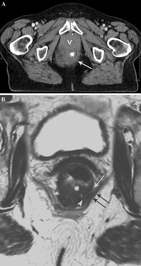
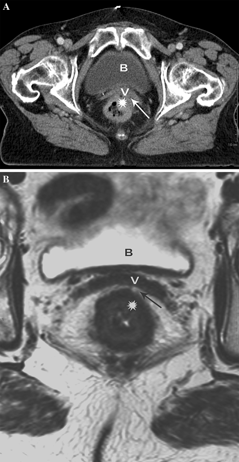
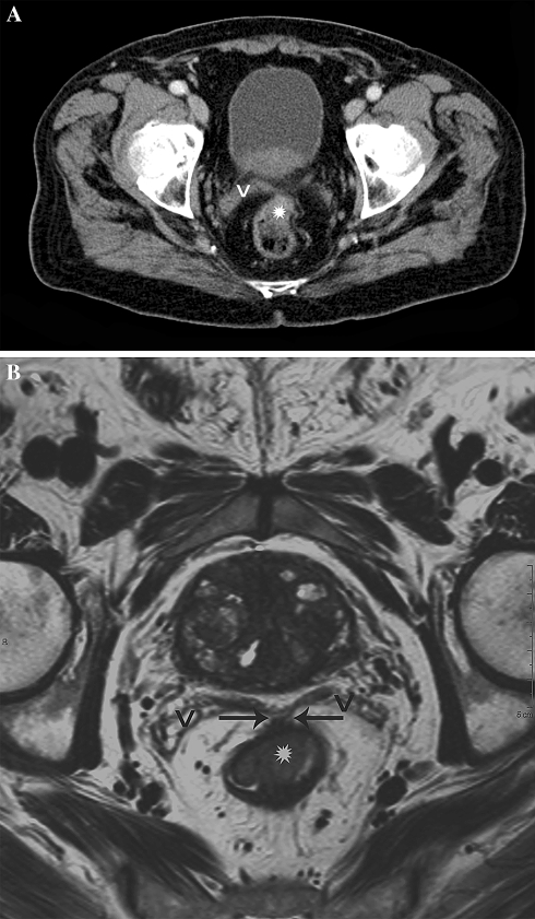
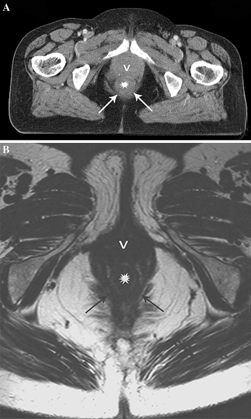
Materials and methods: A total of 35 patients with primary rectal cancer underwent preoperative staging magnetic resonance imaging (MRI) and MDCT. The tumor relationship to the MRF, expressed in 3 categories (1--tumor free MRF = tumor distance > or = 1 mm; 2--threatened = distance < 1 mm; 3--invasion = distance 0 mm) was determined on CT by two observers at patient level and at different anatomical locations. A third expert reader evaluated the MRF tumor relationship on MRI, which served as reference standard. Receiver operating characteristic curves (ROC-curves) and areas under these curves (AUC) were calculated. The inter-observer agreement of CT was determined by using linear weighted kappa statistics.

Results: The AUC of CT for MRF invasion was 0.71 for observer 1 and 0.62 for observer 2. The inter-observer agreement was kappa = 0.34. The performance of CT at mid-high rectal levels was statistically significant better compared to low anterior (obs.1: AUC = 0.88 vs. 0.50; obs 2: AUC = 0.84 vs. 0.31; P < or = 0.040).

Conclusion: Multi-detector row CT has a poor accuracy for predicting MRF invasion in low-anterior located tumors. The accuracy of CT significantly improves for tumors in the mid-high rectum. There is a high inconsistency among readers.


Figures

Fig. 1.
Fig. 1.
Poor anatomical detail on CT leading to overestimation of tumor invasion of the MRF in distal rectal tumors. A Axial MS-CT image of distal rectal cancer. The tumor (asterisk) is difficult to delineate and no fat pad can be seen between the tumor and the pelvic floor (arrow) suggesting invasion of the MRF. B Axial T2-weighted MR image at the same level shows a tumor free MRF represented by a partial intact muscular rectal wall layer (arrowhead) and a minimal fat pad (arrow) inbetween the tumor and the pelvic floor muscles (double arrow).
Fig. 2.
Fig. 2.
Another example of poor anatomical detail on CT causing overestimation of MRF invasion in difficult anatomical regions. A Axial MS-CT image of distal rectal cancer (asterisk) showing tumor invasion of the anterior MRF (arrow) and vagina (v). B Axial T2-weighted MR images at the same level shows a tumor free anterior MRF and vagina (v) represented by a well-appreciated fat pad (arrow) between the tumor (asterisk) and the anterior MRF.
Fig. 3.
Fig. 3.
Difficult visualization of the tumor localization on CT leading to underestimation of tumor invasion of the MRF. A Axial MS-CT image. The rectal tumor located in the anterior rectum (asterisk) at the level of the seminal vesicles (v) is difficult to appreciate due to partial collapse of the rectal lumen and suboptimal soft tissue contrast resolution. B Axial T2-weighted MR image at the same level illustrates an optimal visualization of the tumor in the rectal wall due to high anatomical detail. Also the tumor spread into the anterior MRF (arrows) is well appreciated because of the high soft tissue contrast resolution (V = seminal vesicles).
Fig. 4.
Fig. 4.
Normal rectal wall staged as tumor invasion of the MRF on CT due to insufficient anatomical detail. A Axial MS-CT image suggest a thickened rectal wall interpreted as distal tumor (asterisk) contacting the pelvic floor (arrows) and vagina (v). B Axial T2-weighted MR image at the same level clearly depicts a normal rectal wall (asterisk) as well as surrounding anatomy (arrows = pelvic floor; v = vagina).

References

    1. Heald RJ. Local recurrence of rectal cancer. Dis Colon rectum. 1987;30:572. doi: 10.1007/BF02554794. - DOI - PubMed
    1. Enker WE. Total mesorectal excision-the new golden standard of surgery for rectal cancer. Ann Med. 1997;29:127–133. - PubMed
    1. Kapiteijn E, Marijnen CA, Nagtegaal ID, et al. Preoperative radiotherapy combined with total mesorectal excision for resectable rectal cancer. N Engl J Med. 2001;345:638–646. doi: 10.1056/NEJMoa010580. - DOI - PubMed
    1. Sauer R, Beckers H, Hohenberger W, et al. Preoperative vs postoperative chemoradiotherapy for rectal cancer. N Engl J Med. 2004;351:1731–1740. doi: 10.1056/NEJMoa040694. - DOI - PubMed
    1. Valentini V, Glimelius B, Minsky BD, et al. The multidisciplinary rectal cancer treatment: main convergences, controversial aspects and investigational areas which support the need for an European consensus. Radiother Oncol. 2005;76:241–250. doi: 10.1016/j.radonc.2005.07.001. - DOI - PubMed