. 2008 Feb;4(2):119-25.
doi: 10.1038/nchembio.63. Epub 2008 Jan 6.

A forward chemical genetic screen reveals an inhibitor of the Mre11-Rad50-Nbs1 complex

Affiliations

A forward chemical genetic screen reveals an inhibitor of the Mre11-Rad50-Nbs1 complex

Aude Dupré et al. Nat Chem Biol. 2008 Feb.

Abstract

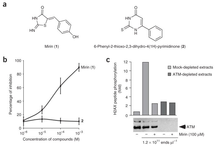
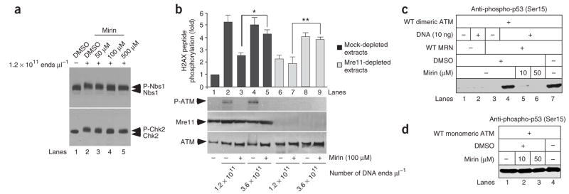
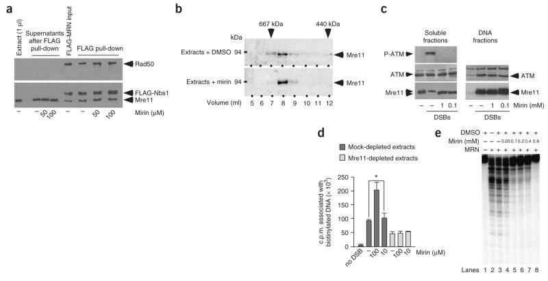
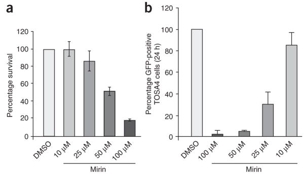
The MRN (Mre11-Rad50-Nbs1)-ATM (ataxia-telangiectasia mutated) pathway is essential for sensing and signaling from DNA double-strand breaks. The MRN complex acts as a DNA damage sensor, maintains genome stability during DNA replication, promotes homology-dependent DNA repair and activates ATM. MRN is essential for cell viability, which has limited functional studies of the complex. Small-molecule inhibitors of MRN could circumvent this experimental limitation and could also be used as cellular radio- and chemosensitization compounds. Using cell-free systems that recapitulate faithfully the MRN-ATM signaling pathway, we designed a forward chemical genetic screen to identify inhibitors of the pathway, and we isolated 6-(4-hydroxyphenyl)-2-thioxo-2,3-dihydro-4(1H)-pyrimidinone (mirin, 1) as an inhibitor of MRN. Mirin prevents MRN-dependent activation of ATM without affecting ATM protein kinase activity, and it inhibits Mre11-associated exonuclease activity. Consistent with its ability to target the MRN complex, mirin abolishes the G2/M checkpoint and homology-dependent repair in mammalian cells.


Figures

Figure 1
Figure 1
Identification of mirin as an inhibitor of ATM activation by DSBs in X. laevis extracts. (a) Structure of mirin (1) and of 6-phenyl-2- thioxo-2,3-dihydro-4(1H)-pyrimidinone (2). (b) Titration curve of mirin and 2 on H2AX peptide phosphorylation assay. X. laevis extracts were incubated with increasing concentrations of either mirin or 2 in the presence of DSBcontaining DNA (5 ng μl−1). ATM activity was assayed as described in Supplementary Figure 1, and percentages of inhibition were calculated as described in Methods. Each bar represents the average of three different experiments with s.d. shown. (c) Mock-depleted extracts (lanes 1 to 3) or ATM-depleted extracts (lanes 4 and 5) were incubated with streptavidin-bound biotinylated DNA (DNA, lanes 2 to 5) in the presence of 100 μM mirin (lanes 3 and 5) or DMSO (lanes 2, 4, 6 and 8). After DNA pull-down, ATM activation was assayed in soluble fractions by measuring H2AX peptide phosphorylation. The western blot below shows the extent of ATM depletion.
Figure 2
Figure 2
Mirin inhibits MRN-dependent activation of ATM. (a) Mirin inhibits ATM-dependent phosphorylation of Nbs1 and Chk2. X. laevis extracts were incubated with DMSO or mirin as indicated and then treated with DSBs. Nbs1 (upper panel) and Chk2 (lower panel) electrophoretic mobility were monitored by western blot. (b) Mirin inhibits MRN-dependent activation of ATM. Mock-treated (dark gray) or Mre11-depleted (light gray) extracts were incubated with 100 μM of mirin (lanes 3, 5, 7 and 9). ATM activation was triggered by addition of DSB-containing DNA at the indicated concentrations. Following DNA pull-down, ATM activation was assayed in the soluble fractions by H2AX peptide phosphorylation, or by western blot for phospho-Ser1981 of ATM (P-ATM). Each bar represents the average of four different experiments with s.d. shown. Values marked with asterisks are significantly different (P < 0.003). Mre11 depletion is shown below the graph. (c) Mirin prevents MRN- and DNA-dependent activation of dimeric ATM in vitro. Dimeric ATM activity was assayed in the presence of DNA (lane 2), MRN (lane 3), MRN and DNA (lanes 4 to 7), and with or without mirin (lanes 5 and 6). ATM activation was monitored by western analysis of Ser15-phosphorylated p53. (d) Mirin does not inhibit the activity of monomeric ATM in vitro. Monomeric ATM activity was assayed as in c in the presence of mirin, as indicated. WT, wild type.
Figure 3
Figure 3
Mirin inhibits the nuclease activity of Mre11. (a) Mirin does not trigger MRN complex dissociation in extracts. Extracts were incubated with FLAG-tagged MRN complex and then incubated with DMSO (−) or mirin. Recombinant MRN was isolated, and supernatants or FLAG-resin were probed using antibodies against FLAG, Mre11 or Rad50. X. laevis Rad50 protein is not recognized by the Rad50 antibody. (b) Mirin does not dissociate endogenous Mre11-associated complexes. Extracts were treated with DMSO or 100 μM mirin and fractionated on a Superose 6 gel-filtration column. Fractions were probed by western blot for Mre11. Molecular weight markers are indicated on top. (c) Mirin does not inhibit ATM and Mre11 binding to DNA. Extracts were treated with DMSO (−) or with mirin before incubation with DSB-containing DNA (1.2 × 10¹¹ ends μl−1). Following DNA pull-down, phosphorylation of ATM Ser1981 (upper panel), ATM (middle panel) and Mre11 (lower panel) were monitored by western blotting in soluble fractions and DNA fractions. (d) Mirin does not inhibit MRN-associated DNA tethering activity. Mock-treated (dark gray) or Mre11-depleted (light gray) extracts were incubated with DMSO (−) or mirin at the indicated concentrations. Extracts were then incubated with streptavidin-bound DNA and free radioactive DNA. DNA-tethering activity was assayed by measuring the radioactivity associated with streptavidin-bound DNA. Each bar represents an average of six independent experiments with s.d. shown. Values marked with asterisks are significantly different (P = 0.001). (e) Mirin inhibits the nuclease activity of MRN. Purified human MRN was incubated with a 5′-labeled double-strand oligonucleotide in the presence of DMSO or mirin, as indicated. Digested DNA products were analyzed by denaturing PAGE followed by autoradiography
Figure 4
Figure 4
Mirin abolishes the G2/M checkpoint. (a) Cell cycle distribution of TOSA4 cells. TOSA4 cells were treated with DMSO or mirin at the indicated concentrations, and cells were processed for flow cytometric analysis. The cell cycle distribution (G1, S and G2/M) is shown. Each bar represents an average of three independent experiments with s.d. shown. (b) Mirin prevents ATM autophosphorylation on Ser1981. U2OS cells synchronized in G1 were incubated with the indicated concentrations of mirin and then mock-irradiated or irradiated with 10 Gy. Cells were harvested 30 min after irradiation and processed for western blot with antibodies against phosphorylated Ser1981 of ATM (top panel) or total ATM (bottom panel). (c) Mirin abolishes the IR-induced G2/M checkpoint in U2OS cells. U2OS cells were synchronized in G2 (see Methods), treated with DMSO (control) or 25 μM mirin, and irradiated with 10 Gy, and mitotic cells were trapped with nocodazole. A typical FACS profile for each sample is shown on top. The average percentage of phospho-H3-positive cells (in mitosis) following irradiation is shown in control and mirin-treated cells in the graph below. The average of three independent experiments with s.d. is shown.
Figure 5
Figure 5
Mirin inhibits homology-dependent DNA repair in human cells. (a) Cytotoxicity of mirin in HEK293 cells. HEK293 cells were treated with the indicated concentrations of mirin, then fixed and stained 10 d later (see Methods). Stained plates were counted for colonies. Percentage survival is expressed as the average number of colonies on treated plates divided by the average number of colonies on control plates (0 μM mirin). Each bar represents an average of three independent experiments with s.d. shown. (b) Gene conversion assay. TOSA4 cells treated with DMSO or mirin at the indicated concentrations were transfected with I-Sce1–expressing plasmid. After 24 h, GFP-expressing cells were then scored by cell sorting. The average of four independent experiments is shown. Each bar represents an average of five independent experiments with s.d. shown.

Comment in

Similar articles

Cited by

References

    1. Zhou BB, Elledge SJ. The DNA damage response: putting checkpoints in perspective. Nature. 2000;408:433–439. - PubMed
    1. Kastan MB, Bartek J. Cell-cycle checkpoints and cancer. Nature. 2004;432:316–323. - PubMed
    1. Lavin MF, et al. ATM signaling and genomic stability in response to DNA damage. Mutat Res. 2005;569:123–132. - PubMed
    1. Bakkenist CJ, Kastan MB. DNA damage activates ATM through intermolecular autophosphorylation and dimer dissociation. Nature. 2003;421:499–506. - PubMed
    1. You Z, Chahwan C, Bailis J, Hunter T, Russell P. ATM activation and its recruitment to damaged DNA require binding to the C terminus of Nbs1. Mol Cell Biol. 2005;25:5363–5379. - PMC - PubMed

Publication types

MeSH terms