. 2007 Aug;2(4):403-422.
doi: 10.2217/17460875.2.4.403.

Role of Lipids in Brain Injury and Diseases

Affiliations

Role of Lipids in Brain Injury and Diseases

Rao Muralikrishna Adibhatla et al. Future Lipidol. 2007 Aug.

Abstract

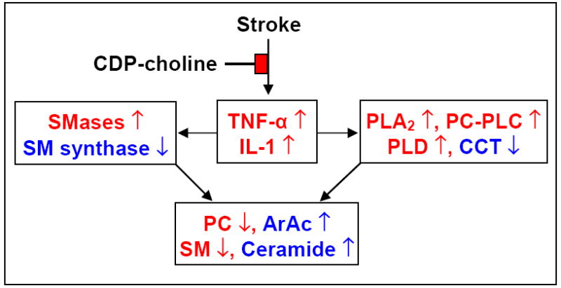
Lipid metabolism is of particular interest due to its high concentration in CNS. The importance of lipids in cell signaling and tissue physiology is demonstrated by many CNS disorders and injuries that involve deregulated metabolism. The long suffering lipid field is gaining reputation and respect as evidenced through the Center of Biomedical Research Excellence in Lipidomics and Pathobiology (COBRE), Lipid MAPS (Metabolites And Pathways Strategy) Consortium sponsored by NIH, European initiatives for decoding the lipids through genomic approaches, and Genomics of Lipid-associated Disorder (GOLD) project initiated by Austrian government. This review attempts to provide an overview of the lipid imbalances associated with neurological disorders (Alzheimer's, Parkinson's; Niemann-Pick; Multiple sclerosis, Huntington, amyotrophic lateral sclerosis, schizophrenia, bipolar disorders and epilepsy) and CNS injury (Stroke, traumatic brain injury; and spinal cord injury) and a few provocative thoughts. Lipidomic analyses along with RNA silencing will provide new insights into the role of lipid intermediates in cell signaling and hopefully open new avenues for prevention or treatment options.


Figures

Figure 1
Figure 1. A) Lipid metabolism in ischemic neuronal death
Activation of phospholipases (PLA2, PC-PLC, PI-PLC and PLD) following cerebral ischemia results in release of lipid second messengers 1,2-diacylglycerol (DAG), phosphatidic acid (PA), lyso-phosphatidic acid (lyso-PA), docosahexaenoic acid (DHA), and arachidonic acid (ArAc). PA and DAG can be readily inter-converted by phosphohydrolases and DAG-kinases. ArAc undergoes further metabolism by cyclooxygenases/lipoxygenases (COX/LOX) to generate important signaling and vasoactive eicosanoids. Free radicals are formed during ArAc metabolism by COX/LOX and free radical generation can be induced by eicosanoids. ArAc generates pro-inflammatory prostaglandins, leukotrienes, and thromboxanes as well as LOX-generated anti-inflammatory lipoxins. Through the LOX pathway, DHA is metabolized to anti-inflammatory resolvins and protectins, including 10,17S-docosatriene (Neuroprotectin D1), an endogenous neuroprotectant. B). Glycerophospholipid and sphingolipid relationship. Tumor necrosis factor-α (TNF-α) and interleukin-1 (IL-1) activate neutral sphingomyelinase (N-SMase) and acidic sphingomyelinase (A-SMase) through stimulation of PLA2 and PC-PLC and release of ArAc and DAG, respectively. N-SMase and A-SMase hydrolyze sphingomyelin (SM) to liberate ceramide. SM synthase transfers the phosphocholine head-group of PC to ceramide to form SM and DAG.
Figure 2
Figure 2. Time course of changes related to cytokines, lipid metabolism and oxidative stress after transient brain ischemia
↑=increase, ↓=decrease, compared to control. ArAc: arachidonic acid; CCT: cytidine triphosphate:phosphocholine cytidylyltransferase; HNE: 4-hydroxynonenal; IL-1β: interleukin-1β; MDA: malondialdehyde; •OH: hydroxyl radical; PLA2: phospholipase A2; PC-PLC: PC-phospholipase C; PLD2: phospholipase D2; PLA2 enzyme activity, sPLA2 mRNA and protein expression, PC-PLC activity and PLD2 protein expression were increased after stroke. CCT catalyzes the rate-limiting step in the biosynthesis of PC. CCT activity and protein expression decreased following stroke. Activation of phospholipases and loss of CCT collectively resulted in loss of PC.
Figure 3
Figure 3. CDP-choline: Integration of cytokine response and lipid metabolism after stroke
[74] Upregulation of TNF-α and IL-1 differentially affects phospholipases, SMases; CCT and SM synthase in collectively causing loss of PC and SM and release of ArAc and ceramide. CDP-choline partially prevents loss of these phospholipids by attenuating the inflammatory response.
Figure 4
Figure 4. CDP-choline and PC synthesis
CCT is down-regulated after stroke, impeding PC synthesis. CDP-choline liposomes deliver the drug intact to the brain, bypassing the CCT step. CCT: cytidine triphosphate:phosphocholine cytidylyltransferase. CPT: CDP-choline:1,2-diacylglycerol cholinephosphotransferase. The Western blot shows loss of CCT protein after stroke.

References

    1. Wenk MR. The emerging field of lipidomics. Nat Rev Drug Discov. 2005;4(7):594–610. ●● An excellent comprehensive review on various aspects of lipidomics and bioinformatics.

    1. Fahy E, Subramaniam S, Brown HA, et al. A comprehensive classification system for lipids. J Lipid Res. 2005;46(5):839–862. ● A comprehensive classification of lipids with a common platform that is compatible with informatics requirements for the hard-core lipid community.

    1. Adibhatla RM, Hatcher JF, Dempsey RJ. Lipids and lipidomics in brain injury and diseases. AAPS J. 2006;8(2):E314–E321. - PMC - PubMed
    1. Adibhatla RM, Hatcher JF. Phospholipase A2, reactive oxygen species, and lipid peroxidation in cerebral ischemia. Free Radic Biol Med. 2006;40(3):376–387. - PubMed
    1. Jiang J, Borisenko GG, Osipov A, et al. Arachidonic acid-induced carbon-centered radicals and phospholipid peroxidation in cyclo-oxygenase-2-transfected PC12 cells. J Neurochem. 2004;90(5):1036–1049. - PubMed

LinkOut - more resources