

Autism and the development of face processing
- PMID: 18176635
- PMCID: PMC2174902
- DOI: 10.1016/j.cnr.2006.08.001
Autism and the development of face processing
Abstract
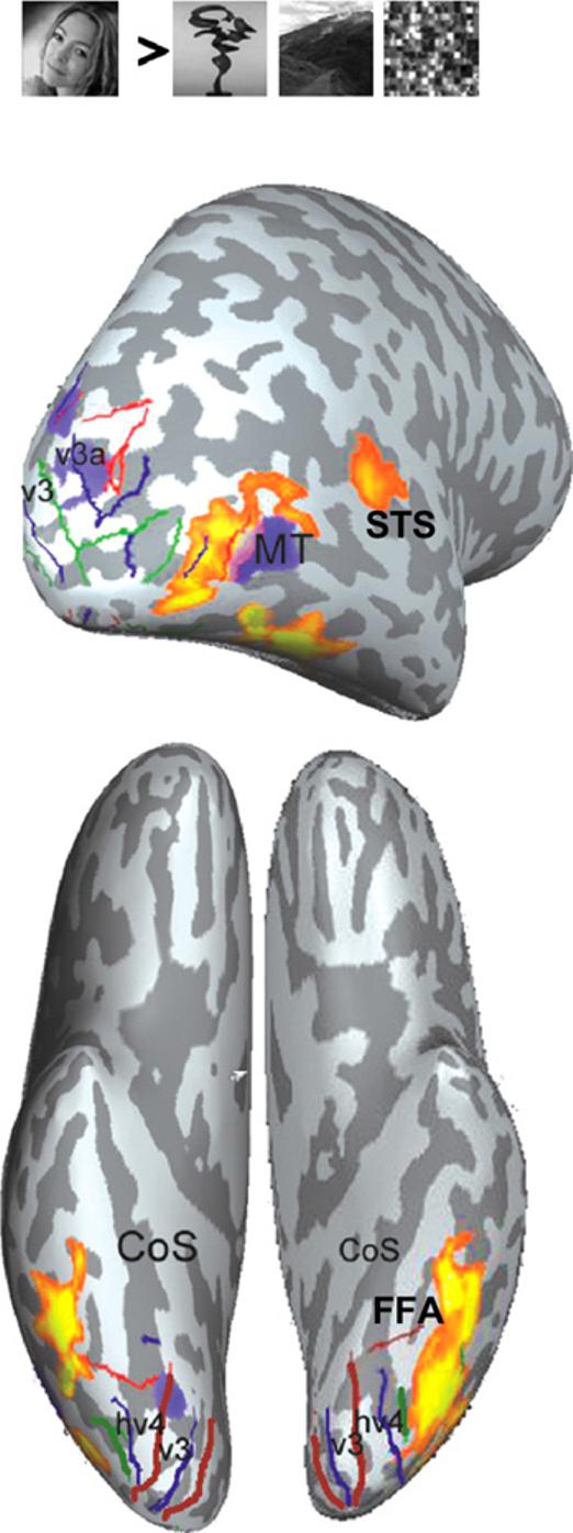
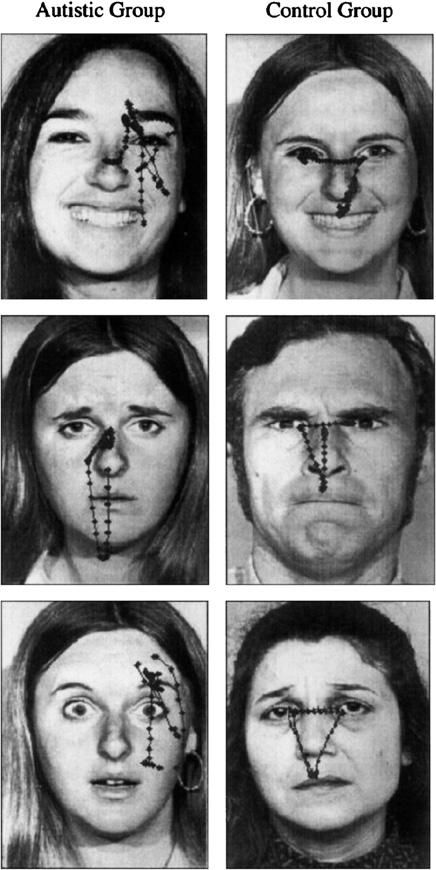
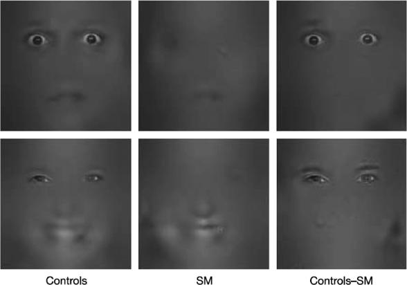
Autism is a pervasive developmental condition, characterized by impairments in non-verbal communication, social relationships and stereotypical patterns of behavior. A large body of evidence suggests that several aspects of face processing are impaired in autism, including anomalies in gaze processing, memory for facial identity and recognition of facial expressions of emotion. In search of neural markers of anomalous face processing in autism, much interest has focused on a network of brain regions that are implicated in social cognition and face processing. In this review, we will focus on three such regions, namely the STS for its role in processing gaze and facial movements, the FFA in face detection and identification and the amygdala in processing facial expressions of emotion. Much evidence suggests that a better understanding of the normal development of these specialized regions is essential for discovering the neural bases of face processing anomalies in autism. Thus, we will also examine the available literature on the normal development of face processing. Key unknowns in this research area are the neuro-developmental processes, the role of experience and the interactions among components of the face processing system in shaping each of the specialized regions for processing faces during normal development and in autism.
Figures
References
-
- Allison T, Puce A, McCarthy G. Social perception from visual cues: role of the STS region. Trends Cogn Sci. 2000;4(7):267–78. - PubMed
-
- Winston JS, et al. Automatic and intentional brain responses during evaluation of trustworthiness of faces. Nat Neurosci. 2002;5(3):277–83. - PubMed
-
- Young AW, et al. Facial expression processing after amygdalotomy. Neuropsychologia. 1996;34(1):31–9. - PubMed
Grants and funding
LinkOut - more resources
Full Text Sources
Other Literature Sources