

Exophytic inflammatory myofibroblastic tumor of the stomach in an adult woman: a rare cause of hemoperitoneum
- PMID: 18176977
- PMCID: PMC2673379
- DOI: 10.3748/wjg.14.136
Exophytic inflammatory myofibroblastic tumor of the stomach in an adult woman: a rare cause of hemoperitoneum
Abstract
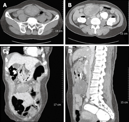
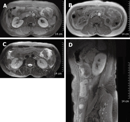
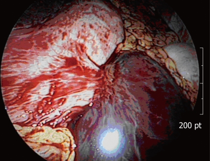
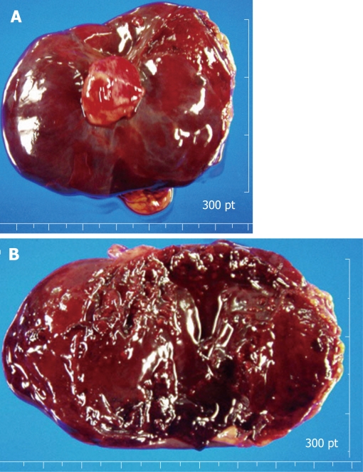
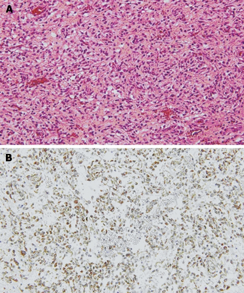
Inflammatory myofibroblastic tumor (IMT) of the stomach in adults is extremely rare, with unpredictable prognosis. We present a 55-year-old woman with a gastric IMT. She experienced sudden abdominal pain 4 d previously. Physical examination showed mild abdominal tenderness in the hypogastrium, but no palpable abnormal abdominal mass. Abdominal CT showed a mass of approximately 8 cm in the gastrocolic ligament. On laparoscopic exploration, unexpected hemoperitoneum of approximately 1.5 L of blood was found, and an exophytic gastric mass of approximately 10 cm, appeared from the anterior wall of the gastric body along the greater curvature. Laparoscopy further showed that non-clotting blood in the abdominal cavity seemed to be from the gastric tumor. After conversion to open surgery for more precise evaluation of the cause of hemoperitoneum and the large friable tumor, gastric wedge resection, including the tumor, was conducted. The final diagnosis was consistent with IMT that originated from the gastric wall.
Figures
References
-
- Coffin CM, Hornick JL, Fletcher CD. Inflammatory myofibroblastic tumor: comparison of clinicopathologic, histologic, and immunohistochemical features including ALK expression in atypical and aggressive cases. Am J Surg Pathol. 2007;31:509–520. - PubMed
-
- Pratap A, Tiwari A, Agarwal B, Pandey SR, Paudel G, Kumar A. Inflammatory myofibroblastic tumor of the abdominal wall simulating rhabdomyosarcoma: report of a case. Surg Today. 2007;37:352–355. - PubMed
-
- Leon CJ, Castillo J, Mebold J, Cortez L, Felmer R. Inflammatory myofibroblastic tumor of the stomach: an unusual complication after gastrectomy. Gastrointest Endosc. 2006;63:347–349. - PubMed
-
- Coffin CM, Patel A, Perkins S, Elenitoba-Johnson KS, Perlman E, Griffin CA. ALK1 and p80 expression and chromosomal rearrangements involving 2p23 in inflammatory myofibroblastic tumor. Mod Pathol. 2001;14:569–576. - PubMed
-
- Yousem SA, Shaw H, Cieply K. Involvement of 2p23 in pulmonary inflammatory pseudotumors. Hum Pathol. 2001;32:428–433. - PubMed
Publication types
MeSH terms
LinkOut - more resources
Full Text Sources
Medical
