. 2008 Jan 3;5(1):e9.
doi: 10.1371/journal.pmed.0050009.

Enhancing exposure of HIV-1 neutralization epitopes through mutations in gp41

Affiliations

Enhancing exposure of HIV-1 neutralization epitopes through mutations in gp41

Catherine A Blish et al. PLoS Med. .

Abstract

Background: The generation of broadly neutralizing antibodies is a priority in the design of vaccines against HIV-1. Unfortunately, most antibodies to HIV-1 are narrow in their specificity, and a basic understanding of how to develop antibodies with broad neutralizing activity is needed. Designing methods to target antibodies to conserved HIV-1 epitopes may allow for the generation of broadly neutralizing antibodies and aid the global fight against AIDS by providing new approaches to block HIV-1 infection. Using a naturally occurring HIV-1 Envelope (Env) variant as a template, we sought to identify features of Env that would enhance exposure of conserved HIV-1 epitopes.

Methods and findings: Within a cohort study of high-risk women in Mombasa, Kenya, we previously identified a subtype A HIV-1 Env variant in one participant that was unusually sensitive to neutralization. Using site-directed mutagenesis, the unusual neutralization sensitivity of this variant was mapped to two amino acid mutations within conserved sites in the transmembrane subunit (gp41) of the HIV-1 Env protein. These two mutations, when introduced into a neutralization-resistant variant from the same participant, resulted in 3- to >360-fold enhanced neutralization by monoclonal antibodies specific for conserved regions of both gp41 and the Env surface subunit, gp120, >780-fold enhanced neutralization by soluble CD4, and >35-fold enhanced neutralization by the antibodies found within a pool of plasmas from unrelated individuals. Enhanced neutralization sensitivity was not explained by differences in Env infectivity, Env concentration, Env shedding, or apparent differences in fusion kinetics. Furthermore, introduction of these mutations into unrelated viral Env sequences, including those from both another subtype A variant and a subtype B variant, resulted in enhanced neutralization susceptibility to gp41- and gp120-specific antibodies, and to plasma antibodies. This enhanced neutralization sensitivity exceeded 1,000-fold in several cases.

Conclusions: Two amino acid mutations within gp41 were identified that expose multiple discontinuous neutralization epitopes on diverse HIV-1 Env proteins. These exposed epitopes were shielded on the unmodified viral Env proteins, and several of the exposed epitopes encompass desired target regions for protective antibodies. Env proteins containing these modifications could act as a scaffold for presentation of such conserved domains, and may aid in developing methods to target antibodies to such regions.


Conflict of interest statement

Competing Interests: The authors have declared that no competing interests exist.

Figures

Figure 1
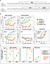
Figure 1. Sequence and Neutralization Profiles of Q461 Viral Variants
(A) The predicted amino acid sequences of the portion of gp41 spanning from the fusion peptide to the MPER of the Q461 viral variants are shown. The locations of HR1, HR2, and MPER are indicated. The Q461e2 sequence is shown as a reference, and the locations of the TA and IV mutations within Q461d1 and the Q461e2 mutants are indicated in the oval and marked with an arrow. A dash indicates no change in the amino acid at that position. (B) Neutralization curves of pseudotyped viruses made with the Env variants described in (A). The y-axis represents the percentage of neutralization, the x-axis represents the concentration of MAb or dilution of plasma tested, as appropriate, and the color and symbol key for the different viral pseudotypes is shown at the bottom. The antibody or plasma sample used is indicated in the inset of the graph, and the global specificity above the charts. The purple line indicates 50% neutralization. (C) The fold-difference in the IC50 values for the Q461e2 mutants and the original neutralization-sensitive Q461d1 variant relative to Q461e2 are shown on log scale, and calculated as described in the Methods. Each point represents the value from a single experiment using an independent viral preparation, for two to four replicate experiments for each virus/antibody combination. Each individual experiment was performed in triplicate. When an IC50 value was not reached for the Q461e2 variant because of its neutralization resistance, the fold-difference for that antibody or plasma sample was calculated using the maximum concentration tested as the IC50 value for Q461e2, and thus reveals the minimum possible difference. A red # sign is marked in the graph above these minimum possible values. The color-coding of the different viruses is shown on the right and is the same as in (B). The specificity of the different antibodies is indicated above the graphs, with different specificities separated by bold lines.
Figure 2
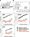
Figure 2. Evaluation of HIV-1 Envelope Concentration and Dissociation of Q461 Envelope Variants
(A) A Western blot of purified viral variants is shown. The viral variant tested is indicated along the top; an equal number of infectious particles was loaded for each viral variant. The “No env” negative control was prepared from cell-free viral supernatant produced from Q23Δenv in the absence of a plasmid encoding a viral Env; because this preparation was unable to infect cells, the maximum possible volume was subjected to purification. The gp160 and gp120 bands are marked with an arrow. (B) Relative gp160 and gp120 expression levels calculated from Western blotting are shown for each of three triplicate experiments. The pseudovirus is indicated on the left, gp120 is shown in the black symbols, and gp160 in the open symbols. Levels of protein expression were normalized relative to the gp160 from Q461d1, which was assigned the level of 100, and marked with an asterisk. The Spearman rank test revealed no correlation between either gp120 or gp160 concentration and susceptibility to any MAb or plasma sample (p > 0.8 for all comparisons). (C) Neutralization curves of purified (closed symbols) and unpurified viral supernatant (open symbols). Results are shown for both Q461d1 (black squares) and Q461e2 (red circles) with 2F5, 4E10, and b12. As in Figure 1B, the y-axis represents the percentage of neutralization, the x-axis represents the concentration of MAb, and the antibody tested is indicated in the inset.
Figure 3
Figure 3. Infectivity and Susceptibility to Enfuviritide of Q461 Envelope Variants
IC50 values (in μg/ml) to enfuviritide of the pseudovirus indicated on the left are shown for each of five (Q461e2.TA, Q461e2.IV, Q461e2.TAIV) or six (Q461d1 and Q461e2) replicate experiments; in some cases points overlap. The Spearman rank test revealed no correlation between the mean enfuviritide concentration and mean susceptibility to any MAb or plasma sample (p > 0.8 for all comparisons).
Figure 4
Figure 4. Sequence, Infectious Titer, and Neutralization Profiles of Q769b9 Envelope Variants
(A) The predicted gp41 amino acid sequences of Q769b9 and the TA, IV, and TAIV mutants. Layout is as described in the legend to Figure 1A. (B) Neutralization curves of pseudotyped viruses made with the Env variants described in (A) are shown. The layout is as described in the legend to Figure 1B. (C) The -fold difference in the IC50 for the Q769b9 mutants relative to Q769b9 is shown on a log scale as in Figure 1C.
Figure 5
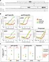
Figure 5. Sequence and Neutralization Profiles of the Subtype B YU-2 Envelope Variants
(A) The predicted gp41 amino acid sequences of YU-2 and the TA, IV, and TAIV mutants. Layout is as described in the legend to Figure 1A. (B) Neutralization curves of pseudotyped viruses made with the Env variants described in (A) are shown. The layout is as described in the legend to Figure 1B. (C) The fold-difference in the IC50 for the YU-2 mutants relative to YU-2 is shown on a log scale as in Figure 1C.

References

    1. Ferrantelli F, Rasmussen RA, Buckley KA, Li PL, Wang T, et al. Complete protection of neonatal rhesus macaques against oral exposure to pathogenic simian-human immunodeficiency virus by human anti-HIV monoclonal antibodies. J Infect Dis. 2004;189:2167–2173. - PubMed
    1. Mascola JR, Lewis MG, Stiegler G, Harris D, VanCott TC, et al. Protection of Macaques against pathogenic simian/human immunodeficiency virus 89.6PD by passive transfer of neutralizing antibodies. J Virol. 1999;73:4009–4018. - PMC - PubMed
    1. Mascola JR, Stiegler G, VanCott TC, Katinger H, Carpenter CB, et al. Protection of macaques against vaginal transmission of a pathogenic HIV-1/SIV chimeric virus by passive infusion of neutralizing antibodies. Nat Med. 2000;6:207–210. - PubMed
    1. Parren PW, Marx PA, Hessell AJ, Luckay A, Harouse J, et al. Antibody protects macaques against vaginal challenge with a pathogenic R5 simian/human immunodeficiency virus at serum levels giving complete neutralization in vitro. J Virol. 2001;75:8340–8347. - PMC - PubMed
    1. Shibata R, Igarashi T, Haigwood N, Buckler-White A, Ogert R, et al. Neutralizing antibody directed against the HIV-1 envelope glycoprotein can completely block HIV-1/SIV chimeric virus infections of macaque monkeys. Nat Med. 1999;5:204–210. - PubMed

Publication types

Associated data