Comparative Study
. 2008 Jun;17(6):489-97.
doi: 10.1111/j.1600-0625.2007.00667.x. Epub 2008 Jan 1.

Estrogen receptor beta expression in nevi during pregnancy

Affiliations
Comparative Study

Estrogen receptor beta expression in nevi during pregnancy

Mary Alice Nading et al. Exp Dermatol. 2008 Jun.

Abstract

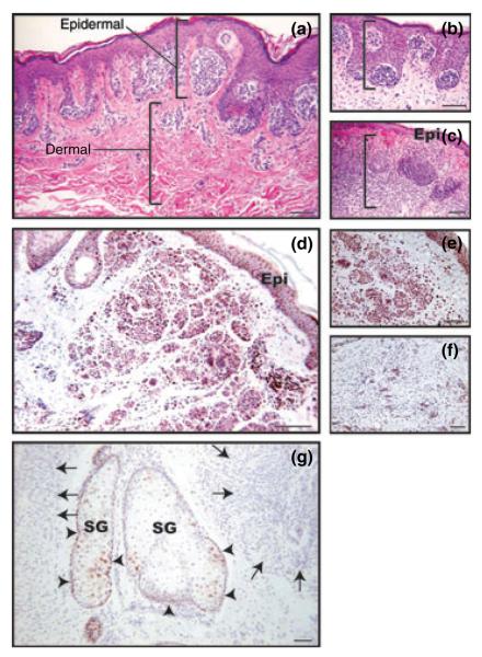
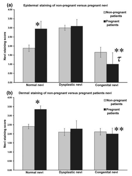
Estrogen levels increase during pregnancy and clinical evidence has long suggested that melanocytes are estrogen-responsive. We hypothesized that nevi from pregnant patients would exhibit increased expression of estrogen receptor beta (ERbeta) and thus enhanced potential to respond to altered estrogen levels. Normal, dysplastic and congenital nevi (n = 212) were collected from pregnant and non-pregnant women ranging from 18 to 45 years of age. Immunohistochemical staining was performed on these nevi using antibodies specifically directed against estrogen receptor alpha (ERalpha) and ERbeta. ERalpha was not observed in any lesions; thus, ERbeta was the predominant estrogen receptor in melanocytic cells from all types of nevi. Enhanced positivity for ERbeta in normal nevi during pregnancy was noted, compared with non-pregnant controls including nevocytes residing in both the epidermal and dermal micro-environments (P = 0.005 and P = 0.001 respectively). Nevi with increasingly melanocytic atypia showed increased ERbeta in nevocytes nested within the epidermis. No additional increase in ERbeta in atypical nevi was observed during pregnancy. For normal and congenital nevi, regardless of pregnancy status, dermally associated nevocytes tended to have greater ERbeta immunoreactivity. Significant decreases in ERbeta immunoreactivity were observed in congenital nevi from pregnant women compared with normal and dysplastic nevi from pregnant women. Our data suggest that nevi possess the capacity to be estrogen-responsive. Factors such as pregnancy and degree of atypia are associated with enhanced ERbeta with the exception of congenital nevi where the melanocytes were unique in their response to pregnancy.


Figures

Figure 1
Figure 1
(a–c) Micrographs stained with H & E illustrate epidermal and dermal regions that were defined for scoring purposes. (a) Dysplastic nevus from the lower back of a 34-year-old non-pregnant woman. The upper bracket shows nests of nevocytes in close association with the epidermis (epidermal nevocytes). The lower bracket shows a few, scattered nevocytes in the dermis (dermal nevocytes). Scale bar = 75 μm. (b) Normal nevus from the neck of a 35-year-old woman. Photo shows the rounded, nest-like morphology that was typical of nevocytes spatially positioned nearest the epidermis (epidermal nevocytes). Bracket indicates the epidermal region. Scale bar = 100 μ. (c) Dysplastic nevus from a 25-year-old woman. Photo shows the scattering of the deep, dermally positioned nevocytes. Bracket indicates spatial location of the dermal nevocytes. Scale bar = 125 μ. (d–f) Micrographs illustrate the spatial differences in morphology and ERβ immunoreactivity of nevocytes from a normal nevus from a 39-year-old non-pregnant patient. (d, e) Show intensely positive cells for ERβ in the upper dermis. Scale bars = 100, 75 μ. (f) In contrast, nevocytes deeply positioned in the dermis are more spindle-like and tend to lose their ERβ expression. Scale bar = 50 μ (g). Photo illustrates negative ERα immunoreactivity in nevocytes (arrows) from a normal nevus from a 43-year-old non-pregnant patient yet ERα-positive nuclear immunostaining is observed in sebocytes adjacent to the lesion (arrowheads). Scale bar = 50 μ. Epi,epidermal layer; SG, sebaceous gland.
Figure 2
Figure 2
Nuclear ERβ nevocyte spatial staining pattern values. (a) Epidermal nevocytes: ERβ staining levels in epidermal nevocytes in lesions in pregnant and non-pregnant patients. Nuclear ERβ levels are significantly higher in normal nevi removed from pregnant patients (*P = 0.0059) when compared with normal nevi from non-pregnant women. Dysplastic nevi showed equally high ERβ levels between the non-pregnant and pregnant patients. Congenital nevi showed a non-statistically significant trend towards a decrease in ERβ in the pregnant patients when compared with non-pregnant women. Congenital nevi during pregnancy showed a decrease in nuclear staining for ERβ when compared with pregnant patients with normal nevi (**P = 0.023). Congenital nevi during pregnancy had a statistically significant decrease in nuclear staining for ERβ when compared with dysplastic nevi from pregnant women (τP = 0.016) Grey columns indicate non-pregnant women. Black columns indicate pregnant patients. Bars indicate the SEM. (b) Dermal nevocytes: ERβ staining levels in dermal nevocytes in lesions in pregnant and non-pregnant patients. Higher staining for ERβ is present in normal nevi from pregnant patients when compared with non-pregnant patients (*P = 0.0014). Congenital nevi during pregnancy showed a decrease in staining for ERβ when compared with pregnant patients with normal nevi (**P = 0.0085).
Figure 3
Figure 3
Immunostaining for ERβ in nevocytic lesions. Normal nevi: (a, c) Photos show immunostaining from a normal nevus from a non-pregnant woman with cell populations showing both positive and negative ERβ immunoreactivity. Scale bars = 100, 50 μ. (b, d) Photos show immunostaining from a normal nevus from a pregnant patient. Greater numbers of immunopositive nevocytes are observed in both the epidermal and dermally positioned nevocytes compared with 3a,c. Scale bars = 100, 50 μ. Congenital nevi: (e, g) Photos show immunostaining in a congenital nevus from the thigh of a 22-year-old non-pregnant woman. Large numbers of intensely positive ERβ nevocytes are prominent (arrows). Scale bars = 200, 50 μ. (f,h) Show immunostaining in a congenital nevus from the breast of a 22-year-old pregnant patient. The numbers of ERβ expressing nevocytes diminish in comparison to non-pregnant patients. Scale bars = 200, 50 μ. Dysplastic nevi: (i, j) show immunostaining in a dysplastic nevus removed from the breast of a 23-year-old non-pregnant patient. Note the high degree of immunoreactivity for ERβ in both the nuclear and cytoplasmic locations. Scale bars = 50, 75 μ. (k, l) Show immunostaining in a dysplastic nevus removed from the breast of a 42-year-old pregnant patient. Note the high degree of immunoreactivity for ERβ in both the nuclear and cytoplasmic locations. Epi, epidermal layer. Scale bars = 50, 75 μ.

References

    1. Elling SV, Powell FC. Physiological changes in the skin during pregnancy. Clin Dermatol. 1997;15:35–43. - PubMed
    1. Kroumpouzos G, Cohen LM. Dermatoses of pregnancy. J Am Acad Dermatol. 2001;45:1–19. quiz 19–22. - PubMed
    1. MacKelfresh J, Chen SC, Monthrope YM. Pregnancy and changes in melanocytic nevi. Obstet Gynecol. 2005;106:857–860. - PubMed
    1. Sanchez JL, Figueroa LD, Rodriguez E. Behavior of melanocytic nevi during pregnancy. Am J Dermatopathol. 1984;6(Suppl):89–91. - PubMed
    1. Zampino MR, Corazza M, Costantino D, Mollica G, Virgili A. Are melanocytic nevi influenced by pregnancy? A dermoscopic evaluation. Dermatol Surg. 2006;32:1497–1504. - PubMed

Publication types

MeSH terms

Substances