. 2008 Apr;18(2):153-63.
doi: 10.1111/j.1750-3639.2007.00107.x. Epub 2008 Jan 3.

Myelin abnormalities without oligodendrocyte loss in periventricular leukomalacia

Affiliations

Myelin abnormalities without oligodendrocyte loss in periventricular leukomalacia

Saraid S Billiards et al. Brain Pathol. 2008 Apr.

Abstract

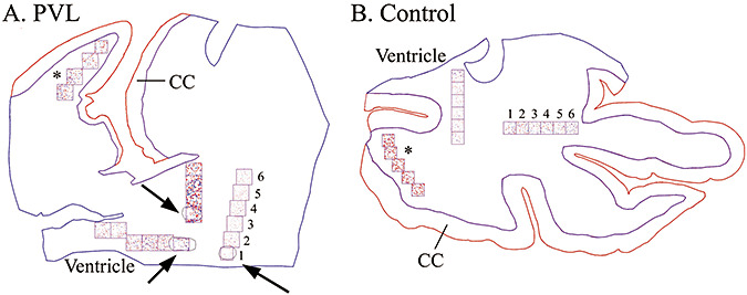
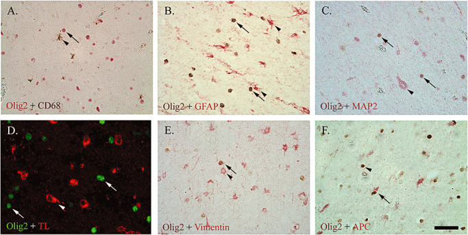
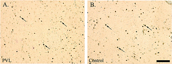
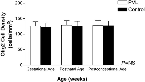
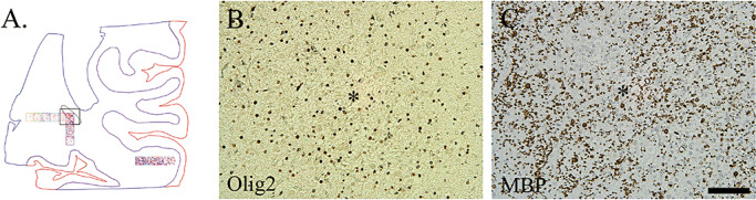
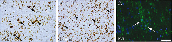
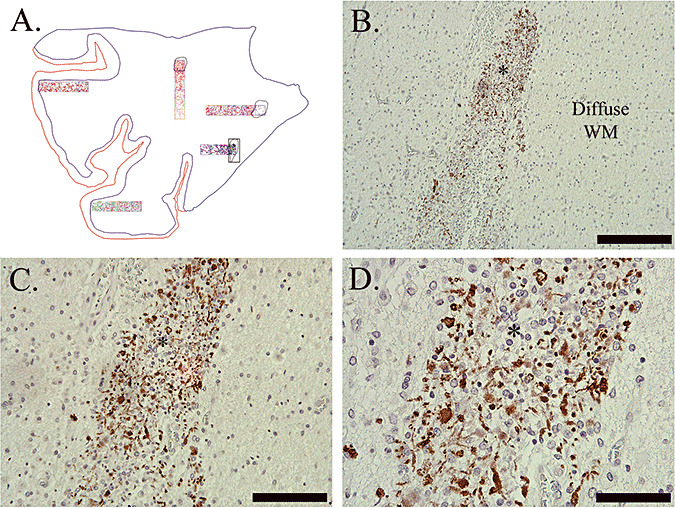
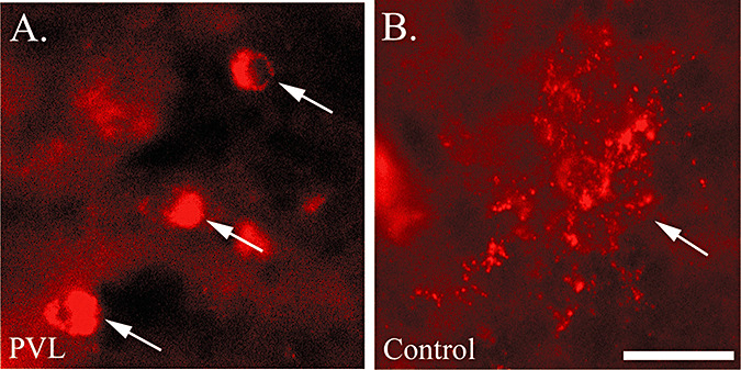
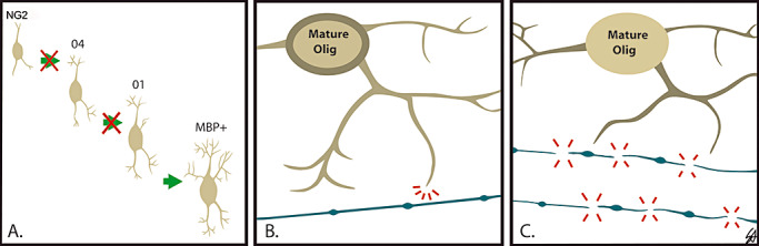
The cellular basis of myelin deficits detected by neuroimaging in long-term survivors of periventricular leukomalacia (PVL) is poorly understood. We tested the hypothesis that oligodendrocyte lineage (OL) cell density is reduced in PVL, thereby contributing to subsequent myelin deficits. Using computer-based methods, we determined OL cell density in sections from 18 PVL and 18 age-adjusted control cases, immunostained with the OL-lineage marker Olig2. Myelination was assessed with myelin basic protein (MBP) immunostaining. We found no significant difference between PVL and control cases in Olig2 cell density in the periventricular or intragyral white matter. We did find, however, a significant increase in Olig2 cell density at the necrotic foci, compared with distant areas. Although no significant difference was found in the degree of MBP immunostaining, we observed qualitative abnormalities of MBP immunostaining in both the diffuse and necrotic components of PVL. Abnormal MBP immunostaining in PVL despite preserved Olig2 cell density may be secondary to arrested OL maturation, damage to OL processes, and/or impaired axonal-OL signaling. OL migration toward the "core" of injury may occur to replenish OL cell number. This study provides new insight into the cellular basis of the myelin deficits observed in survivors of PVL.


Figures

Figure 1
Figure 1
Neurolucida images of Olig2 cell density analysis in a 40 post‐conceptional (PC) week periventricular leukomalacia (PVL) case (A) and a 40 PC week control case (B) (identical counting methods were used for caspase‐3 and Ki67). At low power (20×) the boundaries of the white matter (in blue) and the cerebral cortex (CC) (in red) were outlined. A boundary was then drawn around each necrotic focus (arrows; A). Using a grid system of six 1 mm2 boxes (where possible), with the first box placed over the PVL focus and subsequent boxes progressing away from it, Olig2‐immunopositive cells were counted using pre‐selected markers (at 200×). If more than one necrotic focus was found within each tissue section, a separate analysis was performed, hence the different markers used for each grid system. Analysis of Olig2 cell density was also performed in the distant intragyral white matter (*).
Figure 2
Figure 2
Double‐labeling of Olig2 (nuclear) with the microglial markers CD68 (brown; A) and tomato lectin (TL, red; D), glial fibrillary acidic protein (GFAP)‐positive astrocytes (red; B), microtubule associated protein‐2 (MAP‐2)‐positive neurons (red; C), and vimentin‐positive immature astrocytes (red; E), showed no co‐localization. There was co‐localization, however, between Olig2 and the mature oligodendrocyte lineage (OL) marker Adenomatous Polyposis Coli (APC) (red; F). For images AE, arrows indicate Olig2 and arrow heads indicate other markers. For image F, the arrow indicates the mature OL (Olig2+/APC+), whereas the arrowhead indicates the immature OL (Olig2+/APC−). All images are at 400× magnification. Scale bar = 50 µm.
Figure 3
Figure 3
Olig2 immunostaining (arrows) of nuclei in the diffuse white matter of a 39 post‐conceptional (PC) week periventricular leukomalacia (PVL) case (A) and a 35 PC week control case (B). Images are at 200× magnification. Scale bar = 100 µm.
Figure 4
Figure 4
Olig2 cell density (cells/mm2) (all six boxes), was not different between periventricular leukomalacia (PVL) (n = 18) and control cases (n = 18) adjusting for gestational, post‐natal and post‐conceptional age. Abbreviation: NS = not significant.
Figure 5
Figure 5
A. Neurolucida image of Case 14 (44 post‐conceptional weeks) with a subacute (organizing) necrotic focus (*) located in the optic radiation (highlighted by the black outlined box). Olig2 (B) and myelin basic protein (C) immunostaining were detected within this necrotic focus (*). Images are at 200× magnification. Scale bar = 100 µm.
Figure 6
Figure 6
Increased number of oligodendrocytes (OLs) (observed qualitatively) with myelin basic protein (MBP) immunostaining in the perikaryon (arrows) in periventricular leukomalacia (PVL) cases (A) compared with controls (B) which had more MBP‐positive tubules (arrowheads). Double‐labeling immunofluorescence with MBP (green) and the nuclear marker DAPI (blue) show perikaryl MBP immunostaining (arrows). Images are at 400× magnification. Scale bar = 50 µm.
Figure 7
Figure 7
Abnormal myelin basic protein (MBP) immunostaining in Case 11 (40 post‐conceptional weeks) with a chronic (glial scar) necrotic focus (*). Abnormal MBP immunostaining was in the form of globular segments (lines), rare tubules (arrowhead) and rare sheaths (arrows) (D). Note that the surrounding diffuse white matter (WM) is devoid of any MBP immunostaining (BD). Image A is the Neurolucida image of this case; necrotic focus is highlighted. Image B is taken at 100×, scale = 200 µm; C is at 200×, scale = 100 µm; D is at 400×, scale = 50 µm.
Figure 8
Figure 8
O4 immunostaining of preOLs in a periventricular leukomalacia (PVL) (A) and control (B) case. The morphology of O4+ oligodendrocyte lineages (OLs) in the PVL case lack processes (arrows), whereas in the control case the processes are intact (arrow). Images are taken at 400×, scale = 25 µm.
Figure 9
Figure 9
Proposed hypotheses for the myelin deficits observed in long‐term survivors of periventricular leukomalacia (PVL) in association with preserved Olig2 cell density. A. arrest in the maturation of the oligodendrocyte lineage (OL) progenitor (NG2) to a more mature myelin basic protein‐ (MBP) positive phenotype; B. inability of the mature OL to produce sufficient myelin [note hang‐up of MBP (brown) in the perikaryon]; C. primary axonal injury leading to axonal‐OL communication breakdown.

References

    1. Arai Y, Deguchi K, Mizuguchi M, Takashima S (1995) Expression of beta‐amyloid precursor protein in axons of periventricular leukomalacia brains. Pediatr Neurol 13:161–163. - PubMed
    1. Arnett HA, Fancy SP, Alberta JA, Zhao C, Plant SR, Kaing S et al (2004) bHLH transcription factor Olig1 is required to repair demyelinated lesions in the CNS. Science 306:2111–2115. - PubMed
    1. Back SA, Luo NL, Borenstein NS, Levine JM, Volpe JJ, Kinney HC (2001) Late oligodendrocyte progenitors coincide with the developmental window of vulnerability for human perinatal white matter injury. J Neurosci 21:1302–1312. - PMC - PubMed
    1. Back SA, Luo NL, Borenstein NS, Volpe JJ, Kinney HC (2002) Arrested oligodendrocyte lineage progression during human cerebral white matter development: dissociation between the timing of progenitor differentiation and myelinogenesis. J Neuropathol Exp Neurol 61:197–211. - PubMed
    1. Back SA, Han BH, Luo NL, Chriction CA, Xanthoudakis S, Tam J et al (2002) Selective vulnerability of late oligodendrocyte progenitors to hypoxia‐ischemia. J Neurosci 22:455–463. - PMC - PubMed

Publication types

MeSH terms