. 2007;9(6):224.
doi: 10.1186/ar2333.

Cells of the synovium in rheumatoid arthritis. Macrophages

Affiliations

Cells of the synovium in rheumatoid arthritis. Macrophages

Raimund W Kinne et al. Arthritis Res Ther. 2007.

Abstract

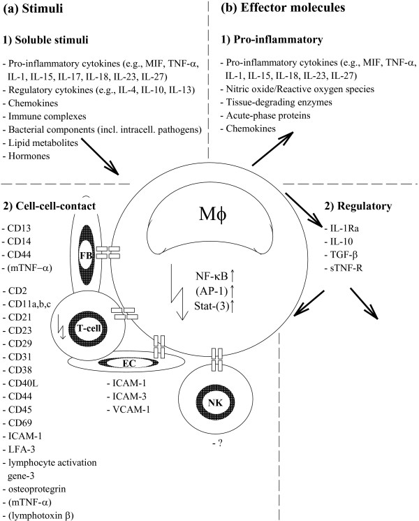
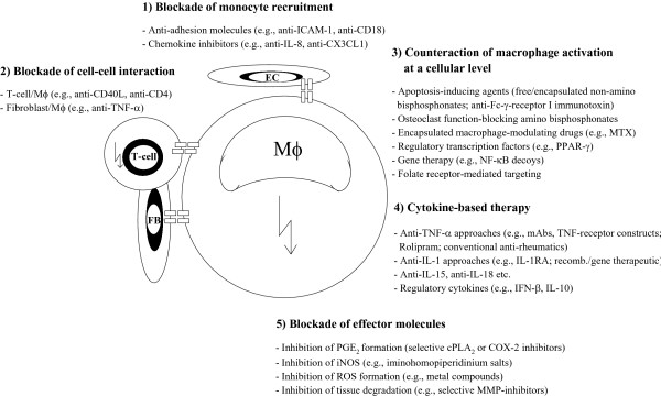
The multitude and abundance of macrophage-derived mediators in rheumatoid arthritis and their paracrine/autocrine effects identify macrophages as local and systemic amplifiers of disease. Although uncovering the etiology of rheumatoid arthritis remains the ultimate means to silence the pathogenetic process, efforts in understanding how activated macrophages influence disease have led to optimization strategies to selectively target macrophages by agents tailored to specific features of macrophage activation. This approach has two advantages: (a) striking the cell population that mediates/amplifies most of the irreversible tissue destruction and (b) sparing other cells that have no (or only marginal) effects on joint damage.


Figures

Figure 1
Figure 1
Physiological/pathological differentiation of the mononuclear phagocyte system in rheumatoid arthritis (RA). (a) Physiological differentiation of the mononuclear phagocyte system (MPS) (steady-state cytokine and growth factor milieu). In the human MPS, monocytes (M) differentiate from a CD34+ stem cell via an intermediate step of monoblasts. Monocytes leave the bone marrow and remain in circulation for approximately 3 days. Upon entering various tissues, they differentiate into different types of resident macrophages (Mφ), including synovial macrophages. It is believed that these mature cells do not recirculate, surviving for several months in their respective tissues until they senesce and die. Some circulating monocytes retain the potential for differentiating into dendritic cells and osteoclasts (asterisk in the insert). The steady-state myeloid differentiation involves many factors, including granulocyte macrophage colony-stimulating factor (GM-CSF), interleukin (IL)-1, IL-6, and tumor necrosis factor-alpha (TNF-α), which are produced by resident bone marrow macrophages (reviewed in [2]). (b) Increased plasticity of myeloid differentiation and its possible role in RA (augmented cytokine and growth factor milieu). Human bone marrow intermediate cells can differentiate into macrophages or dendritic cells in the presence of c-kit ligand, GM-CSF, and TNF-α. TNF-α, in turn, inhibits the differentiation of monocytes into macrophages in vitro and, together with GM-CSF, directs the differentiation of precursor cells into dendritic cells, another important arm of the accessory cell system. Also, either IL-11 or vitamin D3 and dexamethasone induce the differentiation of bone marrow cells or mature macrophages into osteoclasts, cells involved in the destruction of subchondral bone in RA. Osteoclasts and dendritic cells can also be derived from circulating monocytes upon stimulation with macrophage colony-stimulating factor (M-CSF) or IL-4 plus GM-CSF. This plasticity, and its dependence on growth factors or cytokines that are clearly elevated in peripheral blood and bone marrow of patients with RA, may explain some differentiation anomalies in the disease and also the efficacy of some anti-rheumatic drugs. Non-specific enhancement of monocyte maturation and tissue egression, in turn, are consistent with the known alterations in inflammation (reviewed in [2]). The differentiation paths potentially relevant to RA are indicated by bold arrows. The jagged arrows represent possible sites of cell activation. CFU-GM, colony-forming units-granulocyte macrophage; CFU-M, colony-forming units-macrophage; MNC, mononuclear cells; PM(N), polymorphonuclear leukocytes. Reproduced with permission from Kinne RW, Stuhlmuller B, Palombo-Kinne E, Burmester GR: The role of macrophages in rheumatoid arthritis. In Rheumatoid Arthritis. Edited by Firestein GS, Panayi GS, Wollheim FA. New York: Oxford University Press; 2006:55–75 [2].
Figure 2
Figure 2
Paracrine, juxtacrine, and autocrine stimuli (column a) and effector molecules (column b) of macrophage (Mφ) activation in rheumatoid arthritis. Most of the regulatory products of activated macrophages act on macrophages themselves, creating autocrine regulatory loops whose dysregulation possibly promotes disease severity and chronicity. The jagged arrow in the T cell indicates the necessity of pre-activating T cells for effective juxtacrine stimulation of macrophages. AP-1, activation protein; EC, endothelial cells; FB, fibroblasts; ICAM, intracellular adhesion molecule; IL, interleukin; IL-1RA, interleukin-1 receptor antagonist; LFA-3, lymphocyte function-associated antigen-3; MIF, migration inhibitory factor; mTNF-α, mouse tumor necrosis factor-alpha; NF-κB, nuclear factor-kappa-B; NK, natural killer cells; sTNF-R, soluble tumor necrosis factor receptor; TGF-β, transforming growth factor-beta; TNF-α, tumor necrosis factor-alpha; VCAM-1, vascular cell adhesion molecule-1. Reproduced with permission from Kinne RW, Stuhlmuller B, Palombo-Kinne E, Burmester GR: The role of macrophages in rheumatoid arthritis. In Rheumatoid Arthritis. Edited by Firestein GS, Panayi GS, Wollheim FA. New York: Oxford University Press; 2006:55–75 [2].
Figure 3
Figure 3
Potential and established approaches for modulation of monocyte/macrophage (Mφ) functions in rheumatoid arthritis. COX-2, cyclooxygenase-2; EC, endothelial cells; FB, fibroblasts; ICAM-1, intracellular adhesion molecule-1; IFN-β, interferon-beta; IL, interleukin; IL-1RA, interleukin-1 receptor antagonist; iNOS, inducible nitric-oxide synthase; mAbs, mononuclear antibodies; cPLA2, cytosolic phospholipase A2; MMP, metalloprotease; MTX, methotrexate; NF-κB, nuclear factor-kappa-B; PGE2, prostaglandin E2; PPAR-γ, peroxisome proliferator-activated receptor-gamma; ROS, reactive oxygen species; TNF-α, tumor necrosis factor-alpha. Reproduced with permission from Kinne RW, Stuhlmuller B, Palombo-Kinne E, Burmester GR: The role of macrophages in rheumatoid arthritis. In Rheumatoid Arthritis. Edited by Firestein GS, Panayi GS, Wollheim FA. New York: Oxford University Press; 2006:55–75 [2].

Similar articles

Cited by

References

    1. Mulherin D, Fitzgerald O, Bresnihan B. Synovial tissue macrophage populations and articular damage in rheumatoid arthritis. Arthritis Rheum. 1996;39:115–124. doi: 10.1002/art.1780390116. - DOI - PubMed
    1. Kinne RW, Stuhlmuller B, Palombo-Kinne E, Burmester GR. The role of macrophages in rheumatoid arthritis. In: Firestein GS, Panayi GS, Wollheim FA, editor. Rheumatoid Arthritis. New York: Oxford University Press; 2006. pp. 55–75.
    1. Smolen JS, Steiner G. Therapeutic strategies for rheumatoid arthritis. Nat Rev Drug Discov. 2003;2:473–488. doi: 10.1038/nrd1109. - DOI - PubMed
    1. Stuhlmüller B, Ungethüm U, Scholze S, Martinez L, Backhaus M, Kraetsch HG, Kinne RW, Burmester GR. Identification of known and novel genes in activated monocytes from patients with rheumatoid arthritis. Arthritis Rheum. 2000;43:775–790. doi: 10.1002/1529-0131(200004)43:4<775::AID-ANR8>3.0.CO;2-7. - DOI - PubMed
    1. Maeno N, Takei S, Imanaka H, Yamamoto K, Kuriwaki K, Kawano Y, Oda H. Increased interleukin-18 expression in bone marrow of a patient with systemic juvenile idiopathic arthritis and unrecognized macrophage-activation syndrome. Arthritis Rheum. 2004;50:1935–1938. doi: 10.1002/art.20268. - DOI - PubMed

Publication types

MeSH terms