. 2008 Apr 1;44(7):1295-304.
doi: 10.1016/j.freeradbiomed.2007.11.014. Epub 2007 Dec 8.

Reactive oxygen species and oocyte aging: role of superoxide, hydrogen peroxide, and hypochlorous acid

Affiliations

Reactive oxygen species and oocyte aging: role of superoxide, hydrogen peroxide, and hypochlorous acid

Anuradha P Goud et al. Free Radic Biol Med. .

Abstract

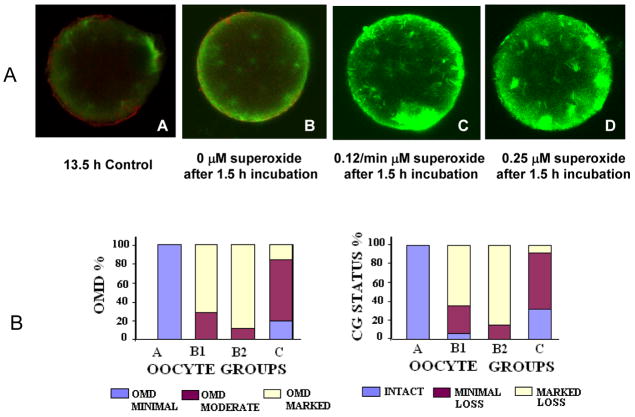
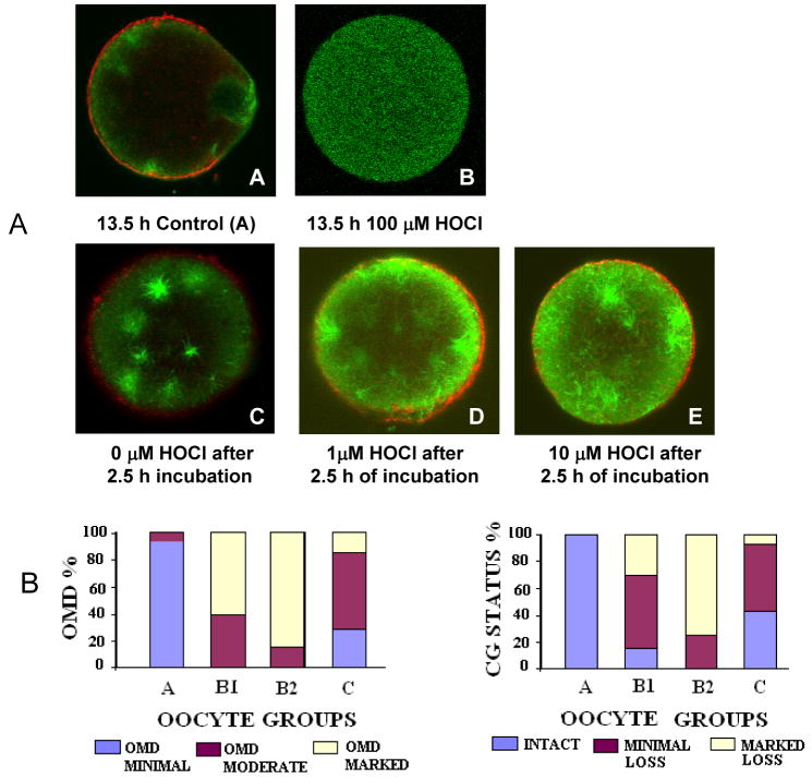
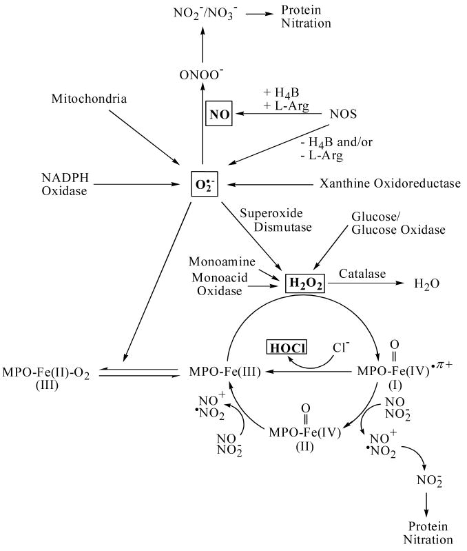
Aging of the unfertilized oocyte inevitably occurs following ovulation, limiting its fertilizable life span. However, the mechanisms that regulate oocyte aging are still unclear. We hypothesize that reactive oxygen species such as superoxide (O2-), hydrogen peroxide (H2O2), and hypochlorous acid (HOCl) are likely candidates that may initiate these changes in the oocyte. In order to test this hypothesis, we investigated direct effects of O2- [hypoxanthine/xanthine oxidase system generating 0.12 (n=42) and 0.25 (n=45) microM O2-/min], H2O2 (20 or 100 microM, n=60), and HOCl, (1, 10, and 100 microM, n=50) on freshly ovulated or relatively old mouse oocytes, while their sibling oocytes were fixed immediately or cultured under physiological conditions (n=96). The aging process was assessed by the zona pellucida dissolution time (ZPDT), ooplasm microtubule dynamics (OMD), and cortical granule (CG) status. The ZPDT increased 2-fold in relatively old, compared to young, untreated oocytes (P<0.0001). Exposure to O2- increased it even further (P<0.0001). Similarly, more O2- exposed oocytes exhibited increased OMD and major CG loss, with fewer having normal OMD and intact CG compared to untreated controls. Interestingly, young oocytes resisted "aging," when exposed to 20 microM H2O2, while the same enhanced the aging phenomena in relatively old oocytes (P<0.05). Exposure to even very low levels of HOCl induced the aging phenomena in young and relatively old oocytes, and higher concentrations of HOCl compromised oocyte viability. Overall, O2-, H2O2, and HOCl each augment oocyte aging, more so in relatively old oocytes, suggesting compromised antioxidant capacity in aging oocytes.


Figures

Fig. 1
Fig. 1
The effect of superoxide generating system on oocyte aging. Panel A shows Fluorescence photomicrographs of oocytes stained for α tubulin (FITC, green) and CG (rhodamine, red) after exposure to hypoxanthine/xanthine oxidase system generating 0.12 and 0.25 μM O2•−/min followed by taxol treatment. An increase in the ooplasmic microtubules is notable in the form of increased free microtubules and asters in panels C (0.12 μM O2•−/min) and D (0.25 μM O2•−/min) compared to the unexposed control oocytes (15 h post hCG, B) and the young controls (13.5 h post-hCG, A), also notable are the total absence of the CG’s in the O2 radical exposed oocytes (C and D) Original magnification: 400–600x; average oocyte diameter ~75–80 μm. Presented in panel B are bar charts showing OMD and CG status among oocytes exposed to the hypoxanthine/xanthine oxidase system generating 0.12 and 0.25 μM O2•−/min. A significantly increased OMD and marked loss in CG is evident in the groups B1 and B2 as compared to the controls A and C. Relatively old oocytes (17.5 h post-hCG) exposed to both 20 and 100 μM H2O2 also showed increased OMD and CG loss (not shown).
Fig. 2
Fig. 2
The effect of H2O2 on oocyte aging. Composite of micrographs shows young oocytes retrieved at 13.5 h exposed to 20 (A–C) or100 μM H2O2 (D–F). Oocytes were stained for α tubulin (FITC, green) and CG (rhodamine, red). An increase in the ooplasmic microtubules is notable in the form of increased free microtubules and asters in the ooplasm (E and F). Pictograph A depicts a young untreated control showing the markedly less OMD and intact CG (D). The young oocytes treated with 20μM H2O2 showed no effect in terms of age related change in OMD (B) and CG (C) as compared to the 100 μM H2O2 exposed oocytes in which OMD is significantly increased (E) and CG is markedly lost (F) as compared to young controls.
Fig. 3
Fig. 3
The effect of hypocholorous acid on oocyte aging. Panel A shows fluorescence photomicrographs of oocytes stained for α tubulin (FITC, green) and CG (rhodamine, red) after exposure to HOCL at 1 μM, 10 μM, 100 μM in PBS followed by taxol treatment. Micrograph A shows the minimal OMD and the intact CG in a young oocyte retrieved at 13.5 h, B shows a typically lysed oocyte fixed and stained immediately after exposure to HOCl (100μM). An increase in the ooplasmic microtubules is notable in the form of increased free microtubules and asters in micrographs D (1 μM) and E (10 μM) compared to the unexposed control oocytes (16.5 h post hCG, C) and the young controls (13.5 h post-hCG, A), also notable are the total absence of the CG’s in the O2•− radical exposed oocytes (C and D). Original magnification: 400–600x; average oocyte diameter ~75–80 μm. Panel B shows the effect of exposure to HOCl at 1 μM, 10 μM, and 100 μM in PBS the min on the OMD and the CG status in the oocytes. A significantly increased OMD and marked loss in CG was seen in the groups B1 and B2 as compared to the controls A and C (P < 0.05).
Fig. 4
Fig. 4
Model showing the factors that modulate the bioavailability of O2•−, H2O2 and HOCl.

Similar articles

Cited by

References

    1. Halliwell B, Gutteridge JMC. In: Free radicals in biology and medicine. Halliwell B, Gutteridge JMC, editors. Oxford: Clarendon Press; 1989. pp. 1–20.
    1. Agarwal A, Allamaneni SS. Role of free radicals in female reproductive diseases and assisted reproduction. Reprod Biomed Online. 2004;9:338–347. - PubMed
    1. Agarwal A, Gupta S, Sharma R. Oxidative stress and its implications in female infertility - a clinician’s perspective. Reprod Biomed Online. 2005;11:641–650. - PubMed
    1. Szczepanska M, Kozlik J, Skrzypczak J, Mikolajczyk M. Oxidative stress may be a piece in the endometriosis puzzle. Ferti l Steril. 2003;79:1288–1293. - PubMed
    1. Van Langendonckt A, Casanas-Roux F, Donnez J. Oxidative stress and peritoneal endometriosis. Fertil Steril. 2002;77:861–870. - PubMed

Publication types