. 2008 Mar 10;314(5):1177-91.
doi: 10.1016/j.yexcr.2007.12.009.

Krp1 (Sarcosin) promotes lateral fusion of myofibril assembly intermediates in cultured mouse cardiomyocytes

Affiliations

Krp1 (Sarcosin) promotes lateral fusion of myofibril assembly intermediates in cultured mouse cardiomyocytes

Cynthia C Greenberg et al. Exp Cell Res. .

Abstract

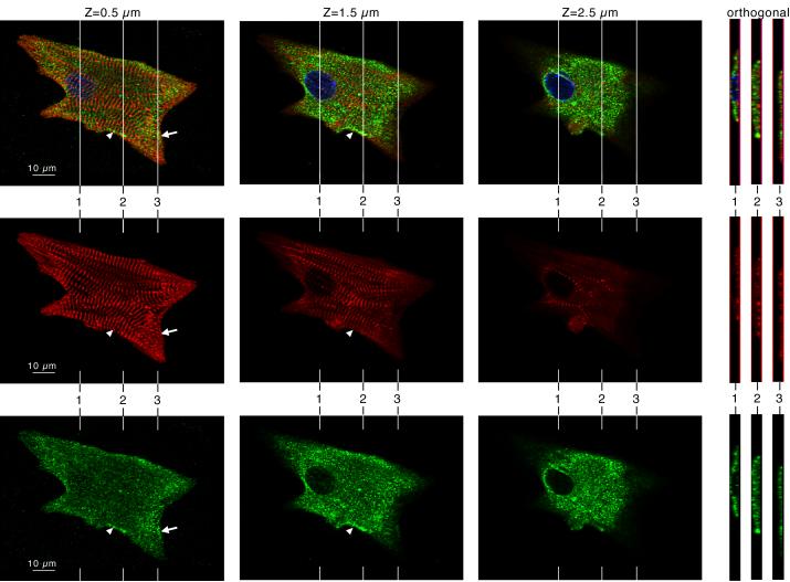
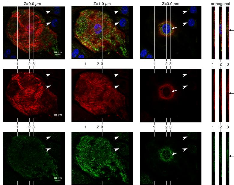
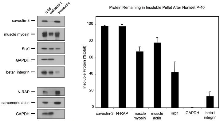
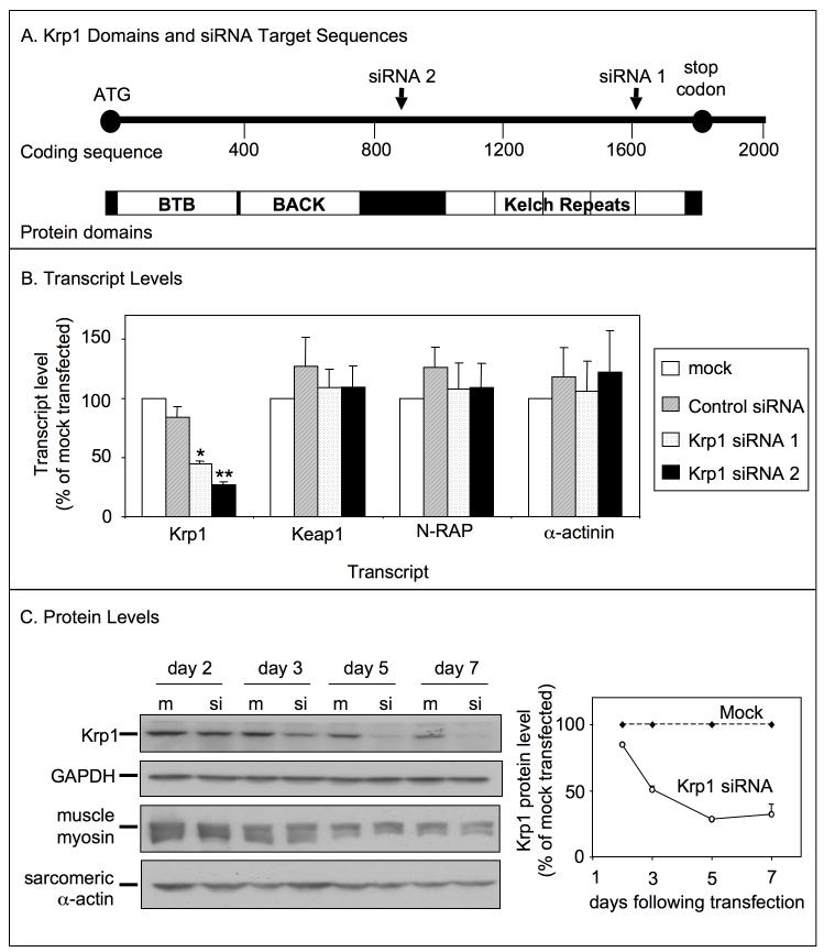
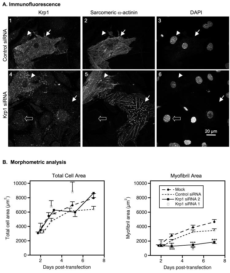
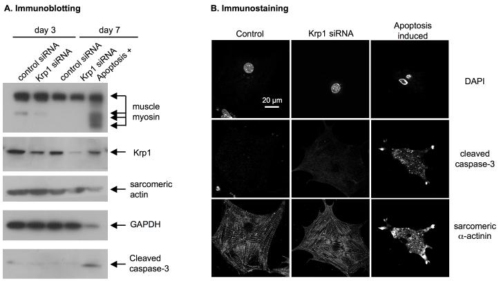
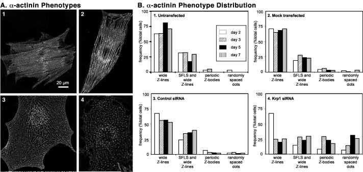
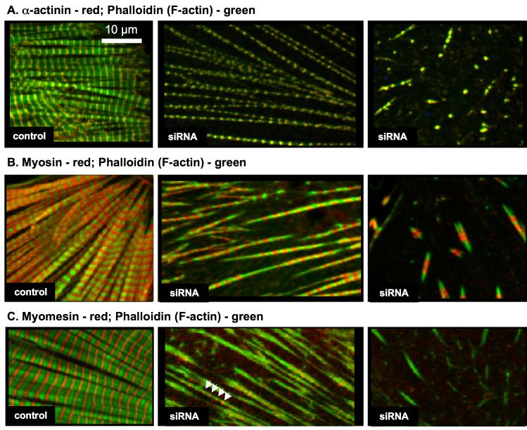
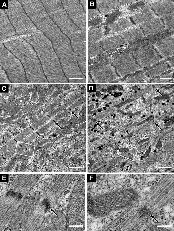
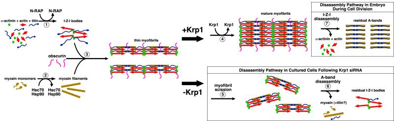
Krp1, also called sarcosin, is a cardiac and skeletal muscle kelch repeat protein hypothesized to promote the assembly of myofibrils, the contractile organelles of striated muscles, through interaction with N-RAP and actin. To elucidate its role, endogenous Krp1 was studied in primary embryonic mouse cardiomyocytes. While immunofluorescence showed punctate Krp1 distribution throughout the cell, detergent extraction revealed a significant pool of Krp1 associated with cytoskeletal elements. Reduction of Krp1 expression with siRNA resulted in specific inhibition of myofibril accumulation with no effect on cell spreading. Immunostaining analysis and electron microscopy revealed that cardiomyocytes lacking Krp1 contained sarcomeric proteins with longitudinal periodicities similar to mature myofibrils, but fibrils remained thin and separated. These thin myofibrils were degraded by a scission mechanism distinct from the myofibril disassembly pathway observed during cell division in the developing heart. The data are consistent with a model in which Krp1 promotes lateral fusion of adjacent thin fibrils into mature, wide myofibrils and contribute insight into mechanisms of myofibrillogenesis and disassembly.


Figures

Figure 1
Figure 1
Krp1 (green, lower row) and α-actinin (red, middle row) localization in a single cultured mouse cardiomyocyte by confocal microscopy. DAPI labeling of the nucleus is also shown in the blue channel of the composite images (top row). Confocal imaging at planes 0.5, 1.5, and 2.5 μm above the adherent ventral surface of the cardiomyocyte are shown, as indicated. Orthogonal views through the cell are shown at the right, and their positions in the x-y plane are indicated in the main micrographs. Most myofibrils lie close to the ventral surface of the cell. Most of the Krp1 lies above the plane of the myofibrils, forming punctate dots in the cytoplasm. Punctate patches of Krp1 are also observed in the plane of the myofibrils, concentrated in premyofibril areas (arrow) as well as alongside narrow myofibrils fusing into more mature structures (arrowhead).
Figure 2
Figure 2
Krp1 (green, lower row) and caveolin-3 (red, middle row) localization in a single cultured mouse cardiomyocyte by confocal microscopy. DAPI labeling of the nucleus is also shown in the blue channel of the composite images (top row). Confocal imaging at planes 0.0, 1.0, and 3.0 μm above the adherent ventral surface of the cardiomyocyte are shown, as indicated. Orthogonal views through the cell are shown at the right, and their positions in the x-y plane are indicated in the adjacent micrographs. The caveolin-3 staining marks the ventral and dorsal surfaces of the cell. Punctate patches of Krp1 are clearly localized within the cytoplasm (arrows), in between the cell boundaries marked by caveolin-3.
Figure 3
Figure 3
Immunoblot analysis of Nonidet P-40 extracted and insoluble fractions. Equal volumes of total cell lysate, extracted and insoluble fractions were analyzed by immunoblot (left) and densitometric analysis (right). Representative immunoblots are shown. The graph displays the means and SEM of three independent experiments. A large proportion of Krp1 remains in the insoluble pellet along with most of the cytoskeletal components (myosin, actin, N-RAP) and caveolin-3, although the majority of Krp1 is present in the extracted supernatant along with the cytosolic marker GAPDH and the integral membrane marker ß1-integrin.
Figure 4
Figure 4
Transfection with siRNA against Krp1 sequences specifically reduces Krp1 expression. (A) Krp1 protein is comprised of an N-terminal BTB-BACK domain and a C-terminal kelch repeat region. In the nucleotide sequence, Krp1 siRNA 1 targets an area within the kelch domain, while Krp1 siRNA 2 targets a sequence between the BTB-BACK and the kelch domains. (B) Transcript levels were analyzed by real time PCR 48 hours after transfection with control or Krp1 siRNA and normalized to levels in mock-transfected controls. Krp1 transcript levels were significantly decreased within 48 hours of transfection with either Krp1 siRNA 1 or 2, while control siRNA had no effect. Reduction of Krp1 expression was specific, as transcript levels of other genes were unchanged. p-values compared to control siRNA-transfected samples: *p<0.05; **p<0.01. Data are the means and SEM of three independent experiments. (C) Immunoblot analysis of specific proteins at the indicated times after mock (m) or Krp1 siRNA (si) transfection. Krp1 protein levels are significantly reduced by day three after transfection, whereas muscle myosin heavy chain and sarcomeric actin levels are unchanged compared to mock-transfected controls. Densitometric analysis of Krp1 protein levels relative to mock-transfected controls is shown on the right; data are the means and SEM from three independent experiments.
Figure 5
Figure 5
Krp1 knockdown decreases mature myofibril content. (A) Cells were fixed and stained for Krp1 and sarcomeric α-actinin 5 days after transfection with control siRNA (top row) or Krp1 siRNA (bottom row). Nuclei were counterstained with DAPI. Images are representative of cells evaluated from three independent cultures. Control cardiomyocytes stain positively for Krp1 and contain sarcomeric α-actinin organized into mature striations (panels 1-3, arrow). Decreased levels of Krp1 after siRNA treatment are associated with low levels of mature myofibrils (panels 4-6, arrow), while some cardiomyocytes retain normal Krp1 levels and exhibit normal striations (panels 4-6, arrowhead). Neighboring fibroblasts exhibit only background staining for Krp1 (panels 1-3, arrowhead; panels 4-6, open arrow). (B) Morphometric analysis of total cardiomyocyte areas (left) and mature myofibril areas (right) versus time. Each point represents the mean and SEM of 25-58 cardiomyocytes from at least 3 independent experiments, with the exception of the Krp1 siRNA 1 data where each point is the mean value of 10-23 cardiomyocytes from 1-3 independent experiments. Krp1 siRNA halts myofibril accumulation after 2 days, but cardiomyocyte spreading is unaffected. p-values compared to mock-transfected controls: ***p <0.001.
Figure 6
Figure 6
Apoptosis is not induced after Krp1 knockdown. Cardiomyocytes were transfected with the indicated siRNA; as a positive control, apoptosis was induced in untreated cells by incubating for 1 day in serum-free, low glucose medium containing 20 mM 2-deoxy-glucose. (A) Immunoblot analysis at 3 and 7 days post-transfection. Krp1 protein levels are decreased after transfection with Krp1 siRNA, but muscle myosin and actin remain at normal levels and cleaved caspase-3 is not detected. (B) Immunofluorescence analysis demonstrates that apoptotic cardiomyocytes display an irregular shrunken morphology with fragmented nuclei and bright, punctate staining for cleaved caspase-3 (right panels). Krp1 siRNA transfected cardiomyocytes display a deficit of mature myofibrils, but their nuclei are intact and caspase-3 levels remain low. Data are representative of two independent experiments.
Figure 7
Figure 7
Classification of cardiomyocytes according to α-actinin organization. Each cardiomyocyte was classified into one of four categories of α-actinin organization corresponding to the dominant phenotype observed by visual inspection. (A) Prototypical examples of α-actinin phenotypes in cultured cardiomyocytes. panel 1: wide Z-lines; panel 2: SFLS and wide Z-lines; panel 3: periodic Z-bodies; panel 4: randomly spaced dots. Panels 1 and 2 are untransfected cells, and cells in panels 3 and 4 were transfected with Krp1 siRNA. After Krp1 siRNA transfection, many cardiomyocytes are almost completely filled with long series of α-actinin dots resembling periodically-spaced Z-bodies or myofibrillogenesis intermediates (panel 3), or are filled with more randomly arranged dots of α-actinin (panel 4). (B) Prevalence of α-actinin phenotypes in cardiomyocytes. Cardiomyocytes were fixed at various times after transfection and analyzed by α-actinin immunostaining and confocal microscopy. Each bar represents the number of cells in the indicated category as a percentage of the total number of cardiomyocytes examined. Panel 1, Untransfected cardiomyocytes: N=120 cardiomyocytes scored from 5 independent experiments. Panel 2, Mock-transfected cardiomyocytes: N=480 cardiomyocytes scored from 10 independent experiments. Panel 3, Control siRNA-transfected cardiomyocytes: N= 355 cardiomyocytes scored from 9 independent experiments. Panel 4, Krp1 siRNA-transfected cardiomyocytes: N=548 cardiomyocytes scored from 11 independent experiments. Cardiomyocytes transfected with Krp1 siRNA were always evaluated in parallel with replicate samples of mock-transfected and/or control siRNA-transfected cells in each experiment.
Figure 8
Figure 8
Organization of sarcomeric components after Krp1 knockdown. Cardiomyocytes were fixed five days post-transfection and double stained with phalloidin to visualize actin filaments (green) and antibodies for either sarcomeric α-actinin (A), muscle myosin (B), or myomesin (C) (red). Examples of control cardiomyocytes (left panels) and cells transfected with Krp1 siRNA 1 (center and right panels) are shown. In control cardiomyocytes, the sarcomeric proteins are organized into mature, well-aligned myofibrils. In contrast, after Krp1 knockdown cells contain sparse, separated fibrils or very short fibrils, which still contain α-actinin, actin and myosin organized in the same banding pattern observed in mature myofibrils. Myomesin is present in the longer fibrils (arrowheads), but is often absent from the shorter fibrils (C, center and right panels, respectively).
Figure 9
Figure 9
Electron micrographs showing examples of myofibrillar structure and organization in control siRNA transfected (A), untransfected (B), and Krp1 siRNA transfected (C-F) cardiomyocytes. Cells were fixed 3 days (A, C, E) or 7 days (B, D, F) after transfection. High magnification micrographs of the boxed areas in (C) and (D) are shown in (E) and (F), respectively. In Krp1 siRNA transfected myocytes, myofibrils are thinner and sparser than in controls. Wide contiguous Z-lines such as in (A) are seldom observed. However, the Z-lines (arrows), thick filaments and thin filaments are organized into normal sarcomeres, and the space between myofibrils remains filled with organelles such as mitochondria, polyribosomes and endoplasmic reticulum that appear normal in structure. Arrowheads in (D) indicate abnormally long Z-lines that further suggest immature myofibrillar structure. The myocyte shown in (D) also has numerous dense membrane-bound granules 100-200 nm in diameter, resembling atrial natriuretic peptide secretory granules. Scale bars = 1 μm (A-D) and 0.2 μm (E, F).
Figure 10
Figure 10
A putative pathway for myofibril assembly (steps 1-4) highlighting the role of transiently associated proteins in organizing the major structural components. Disassembly pathways after Krp1 knockdown (steps 5-6) and during cell division in embryonic development (step 7) are also illustrated. (1) N-RAP promotes assembly of the I-Z-I structures containing actin, α-actinin, and N-terminal titin. (2) Myosin filaments form separately, with appropriate folding and assembly promoted by the Hsc70 and Hsp90 chaperone proteins. (3) Obscurin plays a role in promoting integration of the thick filaments with the I-Z-I structures, with titin associating with the myosin filaments along their length. This gives rise to thin myofibrils. (4) Finally, Krp1 promotes their lateral fusion to form mature myofibrils. (5-6) Krp1 knockdown results in scission of the thin myofibrils, followed by disassembly of the A-bands. The last organized structures observed contain α-actinin and actin, but not myosin. (7) In contrast, disassembly during cell division occurs by removal of α-actinin and actin, leaving organized A-bands. Subsequent A-band disassembly is not shown. See text for details.

Similar articles

Cited by

References

    1. Adams J, Kelso R, Cooley L. The kelch repeat superfamily of proteins: propellers of cell function. Trends Cell Biol. 2000;10:17–24. - PubMed
    1. Stogios PJ, Prive GG. The BACK domain in BTB-kelch proteins. Trends Biochem Sci. 2004;29:634–637. - PubMed
    1. Prag S, Adams JC. Molecular phylogeny of the kelch-repeat superfamily reveals an expansion of BTB/kelch proteins in animals. BMC Bioinformatics. 2003;4:42. - PMC - PubMed
    1. Furukawa M, Xiong Y. BTB protein Keap1 targets antioxidant transcription factor Nrf2 for ubiquitination by the Cullin 3-Roc1 ligase. Mol Cell Biol. 2005;25:162–171. - PMC - PubMed
    1. Cullinan SB, Gordan JD, Jin J, Harper JW, Diehl JA. The Keap1-BTB protein is an adaptor that bridges Nrf2 to a Cul3-based E3 ligase: oxidative stress sensing by a Cul3-Keap1 ligase. Mol Cell Biol. 2004;24:8477–8486. - PMC - PubMed

Publication types