. 2008 Jan 30;26(5):601-13.
doi: 10.1016/j.vaccine.2007.11.084. Epub 2007 Dec 26.

TLR ligands that stimulate the metabolism of vitamin D3 in activated murine dendritic cells can function as effective mucosal adjuvants to subcutaneously administered vaccines

Affiliations

TLR ligands that stimulate the metabolism of vitamin D3 in activated murine dendritic cells can function as effective mucosal adjuvants to subcutaneously administered vaccines

Elena Y Enioutina et al. Vaccine. .

Abstract

Cathelicidin production by human myeloid cells stimulated through toll-like receptor (TLR) 2/1, the migration of human CD8+ T cells to inflamed skin sites, and the ability of murine dendritic cells (DCs) to migrate from skin sites of vaccination to mucosal lymphoid organs all occur via calcitriol-dependent mechanisms. Herein, we report that murine DCs exposed to TLR3/TLR4 ligands upregulate their expression of 1 alpha-hydroxylase, the enzyme that converts circulating 25(OH)D3 to calcitriol, the active form of vitamin D3. TLR3/TLR4 ligands injected subcutaneously affect DC migration in vivo, allowing their trafficking to both draining and non-draining systemic and mucosal lymphoid organs. Subcutaneously delivered vaccines containing TLR3/TLR4 ligands and antigen stimulate the induction of both systemic and mucosal immune responses. Vaccines containing TLR9 ligands fail to stimulate 1 alpha-hydroxylase protein expression, are incapable of redirecting DC migration into Peyer's patches and do not induce mucosal immune responses. These findings support a hypothesis that active metabolites of vitamin D3 produced locally are able to affect various aspects of innate and acquired immune responses.


Figures

Figure 1
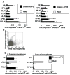
Figure 1. The activation-induced upregulation of 1α-hydroxylase in murine dendritic cells alters their trafficking properties in vivo and their chemotaxis in vitro
(A) CD11c+ BMDCs were stimulated with LPS (10ng/ml) for 24 hours at 37°C or left untreated. Cell lysates were then prepared and analyzed for the presence of 1α-hydroxylase by Western Blot. Proteins were visualized by ECL Plus Western Blotting Detection System. The blot was later stripped and reprobed with antibodies against β-actin to ensure an equal loading of protein. Results are representative of five experiments. (B) CD11c+ BMDCs were matured in vitro for 24 hours in the presence of LPS (10ng/ml) with or without 1α25(OH)2D3 (VD3, 10−8 M) or 25(OH)D3 (10−7 M). The BMDCs were then washed and evaluated for chemotactic responses toward CCL21 (100ng/ml) and S1P (1000nM) using Transwells (5μm pores). After a 3-hour incubation at 37°C, cells that migrated to the bottom chamber were collected and counted. Results are presented as mean of triplicates ± SD. *- difference between numbers of LPS matured DCs in the presence of LPS in the presence of 25(OH)D3 or 1α25(OH)2D3 that migrated into the bottom chamber and numbers of LPS only treated BMDCs that migrated to the bottom chamber was statistically significant (p<0.02-0.009). This experiment was repeated twice with similar results. (C) CD11c+ BMDCs were treated with 10ng/ml of LPS in the presence or absence of 10 8M 1α25(OH)2D3 or 10−7M 25(OH)D3. After a 24-hour incubation at 37°C, the BMDCs were stained with CFSE and subcutaneously injected into naive recipients (three mice per group). Forty-eight hours later, mice were sacrificed and single cell suspensions from individual lymphoid organs (popliteal (PLN), axillary (ALN) lymph nodes, spleens (SPL) and Peyer’s patches (PP)) were prepared and analyzed by FACScan. A total of 400,000 events were collected. Results are presented as mean ± SD. * - difference between numbers of CFSE+ DCs detected in various secondary lymphoid organs of C3H/HeN mice that received a subcutaneous injection of BMDCs exposed to the influences of LPS in the presence of 25(OH)D3 or 1α25(OH)2D3 and numbers of CFSE+ DCs detected in various secondary lymphoid organs of mice that received a subcutaneous injection of BMDCs treated LPS alone was statistically different (p<0.05-0.01).
Figure 2
Figure 2. Dendritic cells leaving a subcutaneous site of LPS injection are capable of trafficking to multiple secondary lymphoid organs throughout the body
(A) Green fluorescent latex microspheres (0.2μm) were injected into the right hind footpads of mature adult C3H/HeN mice in the presence or absence of 1α25(OH)2D3 (0.1μg) or LPS (10μg). Red fluorescent latex microspheres were injected alone into the left hind footpad of the same animals. After forty-eight hours, individual lymphoid tissues were analyzed for the presence of microsphere+ DCs. A total of 400,000 events were collected. Data is presented as mean ± SD. *- differences in the numbers of green microsphere+ DCs in a particular lymphoid organ from animals exposed to the influences of LPS or 1α25(OH)2D3 and the numbers of red microsphere+ DCs was statistically significant (p<0.01-0.003). Results are representative of five independent experiments. (B) Mature adult C3H/HeN mice were injected with 0.2μm green microspheres into the right thigh and 0.2μm red microspheres into the right abdominal area. Both injection sites drain into the same inguinal LN. Forty-eight hours post injection, the draining inguinal LN was collected and single cell suspensions were prepared. Samples were analyzed by FACScan for the presence of green or red microspheres+ DCs. A total of 400,000 events were collected. Experiment was repeated three times with similar results. (C) Green fluorescent microspheres (1.0μm or 0.2μm) were injected in the presence of LPS (10μg) into the right hind footpads of normal mice (three animals per group). Red fluorescent microspheres (1.0μm or 0.2μm) were injected alone into the left hind footpads of the same mice. After forty-eight hours, individual lymphoid organs were removed (popliteal lymph node (PLN), Peyer’s patch (PP)) and single cell suspensions were prepared. Samples were analyzed by FACScan for the presence of microsphere+ DCs. Data presented as mean ± SD. *- difference between numbers of green microsphere+ DCs found in PLN and PPs of animals that received 1.0μm (or 0.2μm) microspheres plus LPS and numbers of red microsphere+ DCs in PLN and PPs of mice that were injected with 1.0μm (or 0.2μm) microspheres alone was statistically significant (p<0.02-0.007). Results are representative of two experiments.
Figure 3
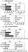
Figure 3. Mobilized antigen-laden DCs that traffic to non-draining lymphoid organs are fully capable of productively initiating an antigen-specific immune response
(A) BALB/c mice received 2×10⁶ CD4+ T cells from DO11.10 donors. After a 24-hour rest, the DO11.10 T cell recipients were subcutaneously immunized with 50μg OVA in Alum in the presence or absence of 0.1μg 1α25(OH)2D3 or 10μg LPS. Recipients were sacrificed 72 hours post-immunization and their secondary lymphoid organs were analyzed for KJ1-26+CD69+ T cells. Data presented as mean ± SD. *- difference between the percentage of KJ1-26+ cells co-expressing CD69 in tested lymphoid organs of animals immunized with OVA and added LPS or 1α25(OH)2D3 and the percentage of KJ1-26+CD69+ cells detected in various lymphoid organs of animals immunized with OVA alone was statistically different (p<0.04-0.02). The results presented are representative of two independent experiments. (B) BALB/c recipients of DO11.10 CD4+ T cells stained with CFSE were subcutaneously immunized with vaccine formulations containing 50μg OVA in Alum with or without 0.1μg 1α25(OH)2D3 or 10μg LPS. After 7 days, individual secondary lymphoid organs were removed, and single cell suspensions were prepared for analysis by FACScan. Data presented as mean ± SD. *- difference between percentage of CFSE+CD4+ cells in various lymphoid organs of mice immunized with OVA plus LPS or 1α25(OH)2D3 and percentage of CFSE+CD4+ cells detected in various lymphoid organs of animals immunized with OVA alone was statistically different (p<0.05-0.01). Results were obtained from two independent experiments.
Figure 4
Figure 4. TLR ligands that upregulate the expression of 1α-hydroxylase in DCs are able to alter their migratory properties following activation-induced mobilization
(A) CD11c+ BMDCs were activated with CpG ODN (20μg/ml), poly I:C (20μg/ml), LPS (10ng/ml) or left untreated. Cells were harvested 24 hours post activation, lysed and analyzed for 1α-hydroxylase protein expression by Western Blot analysis. The blot was later stripped and reprobed with antibodies against β-actin to ensure an equal loading of protein. Results presented are representative of three experiments. (B-D) Green fluorescent microspheres (0.2μm) in the presence of (B) 20μg poly I:C, (C) 10μg LPS, or (D) 20μg CpG ODNs were injected into the right hind footpads of C3H/HeN mice (three per group). Red fluorescent microspheres (0.2μm) were then injected alone into the left hind footpads of the same animals. After forty-eight hours, individual lymphoid organs (right popliteal (RPLN), left popliteal (LPLN), axillary (ALN), cervical (CLN) and mesenteric (MLN) lymph nodes, spleens (SPL) and Peyer’s patches (PP)) were removed. Single cell suspensions were prepared and analyzed by FACScan. Data presented as mean ± SD. *- difference between numbers of green microsphere+ DCs in animals co-injected with microspheres plus poly I:C, LPS or CpG ODN found in various secondary lymphoid organs and numbers of red microsphere+ DCs in the lymphoid organs analyzed from the same mice was statistically significant (p<0.01-0.003). The data presented in Figure 4B–D are representative of three independent experiments.
Figure 5
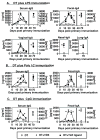
Figure 5. Vaccine formulations, containing LPS or poly I:C as adjuvants are able to promote the induction of both systemic and mucosal immune responses
Groups of mature adult C3H/HeN mice (five per group) were subcutaneously immunized with a vaccine formulation containing 1μg diphtheria CRM 197 protein (DT) and added (A) LPS (10μg), (B) poly I:C (20μg) or (C) CpG ODN (20μg). With each of the TLR ligands being tested, parallel groups were added that were immunized with DT alone, or with DT and 0.1μg 1α25(OH)2D3. Nine weeks after immunization, all groups of animals were subcutaneously reimmunized as described above. Serum and mucosal samples were collected at various time points post vaccination and analyzed for the presence of DT-specific antibodies by ELISA. Results are reported as the mean value of anti-DT antibodies detected in the serum and/or mucosal secretions of five mice per group (±SD). *- difference between levels of anti-DT antibodies in serum or mucosal secretions of mice immunized with DT in the presence of 1α25(OH)2D3 or TLR ligand and levels of anti-DT antibodies in serum or mucosal secretions of mice immunized with DT only were statistically significant (p< 0.004-0.001)

Similar articles

Cited by

References

    1. Takeda K, Kaisho T, Akira S. Toll-like receptors. Annu Rev Immunol. 2003;21:335–76. - PubMed
    1. Smiley ST, King JA, Hancock WW. Fibrinogen stimulates macrophage chemokine secretion through toll-like receptor 4. J Immunol. 2001;167(5):2887–94. - PubMed
    1. Su SL, Tsai CD, Lee CH, Salter DM, Lee HS. Expression and regulation of Toll-like receptor 2 by IL-1beta and fibronectin fragments in human articular chondrocytes. Osteoarthritis Cartilage. 2005;13(10):879–86. - PubMed
    1. Termeer C, Benedix F, Sleeman J, Fieber C, Voith U, Ahrens T, et al. Oligosaccharides of Hyaluronan activate dendritic cells via toll-like receptor 4. J Exp Med. 2002;195(1):99–111. - PMC - PubMed
    1. Biragyn A, Ruffini PA, Leifer CA, Klyushnenkova E, Shakhov A, Chertov O, et al. Toll-like receptor 4-dependent activation of dendritic cells by beta-defensin 2. Science. 2002;298(5595):1025–9. - PubMed

Publication types

MeSH terms