. 2008 Jan 15;105(2):443-8.
doi: 10.1073/pnas.0711075105. Epub 2008 Jan 4.

Nanoelectrochemistry of mammalian cells

Affiliations

Nanoelectrochemistry of mammalian cells

Peng Sun et al. Proc Natl Acad Sci U S A. .

Abstract

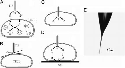
There is a significant current interest in development of new techniques for direct characterization of the intracellular redox state and high-resolution imaging of living cells. We used nanometer-sized amperometric probes in combination with the scanning electrochemical microscope (SECM) to carry out spatially resolved electrochemical experiments in cultured human breast cells. With the tip radius approximately 1,000 times smaller than that of a cell, an electrochemical probe can penetrate a cell and travel inside it without apparent damage to the membrane. The data demonstrate the possibility of measuring the rate of transmembrane charge transport and membrane potential and probing redox properties at the subcellular level. The same experimental setup was used for nanoscale electrochemical imaging of the cell surface.


Conflict of interest statement

The authors declare no conflict of interest.

Figures

Fig. 1.
Fig. 1.
Schematic diagrams of the SECM experiments with single cells (A–D) and an optical micrograph of a typical nanotip used in such experiments (E). (A) The tip is positioned in the solution close to the cell surface. Positive feedback is due to bimolecular electron transfer between hydrophobic redox mediator (O/R) and cell-bound redox moieties (O2/R2). (B) The lipid cell membrane is impermeable for a hydrophilic redox mediator. Negative feedback is due to the hindered diffusion of redox species to the tip electrode. (C) Nanoelectrode voltammetry inside the cell. (D) Positive feedback is produced by mediator regeneration by way of electron transfer at the underlying Au surface.
Fig. 2.
Fig. 2.
Current vs. distance curve obtained with a Pt tip approaching and penetrating an MCF-10A cell (A) and voltammograms obtained at nanoelectrodes inside living cells and in solution (B–D). (A) Solid line is the theoretical curve for an insulating substrate (28). The tip current is normalized by the value measured in the bulk solution. The approach speed was 20 nm/s. (B) Voltammograms obtained in the bulk solution outside of the cell (curve 1), inside the cell (curve 2), and in external solution after the tip was withdrawn from the cell (curve 3). (Inset) Voltammograms obtained in PBS (blue) and inside the cell (pink) without purging the solution with nitrogen. (C) Intracellular voltammograms obtained by scanning the electrode potential between 0 and +0.8 V (Left) and 0 and −0.8 V (Right). The scan rate was (in mV/s): 50 (blue), 20 (red), and 10 (black). The concentration of Ru(NH3)6Cl3 in PBS was 1 mM (A–C) and 0.1 mM (B Inset). (D) Dependence of the steady-state diffusion limiting current vs. concentration of Ru(NH3)6Cl3. The tip radius (in nm) was: 42 (A), 39 (B), 54 (C), and 73 (D).
Fig. 3.
Fig. 3.
Approach and penetration of an MCF-10A cell by a 144-nm tip in PBS solution containing 1 mM FcCH2OH (A) and steady-state voltammograms obtained at the same tip outside (curve 1) and inside (curve 2) the cell (B). (A Inset) Shown is the fit between the experiment (symbols) and the theory (solid line) for the final part of the curve, where the tip approaches the bottom of the Petri dish.
Fig. 4.
Fig. 4.
Voltammograms of FcCH2OH at a 112-nm Pt tip obtained in the bulk solution (curve 1) and inside the same cell (curves 2–5) (A), and corresponding dependences of i(E1/2) vs. cFcCH2OH (B) and ΔE1/2 vs. i(E1/2) (C). (A) cFcCH2OH (in mM) was: 1 (curves 1 and 2), 0.5 (curve 3), 0.25 (curve 4), and 0.125 (curve 5).
Fig. 5.
Fig. 5.
Evaluating the rate of charge/mass transport across the cell membrane from SECM approach curves. The tip radius (in nm) was: 790 (curve 1), 280 (curve 2), and 146 (curve 3). Experimental data (symbols) was fitted to the theory (solid lines) for finite charge transport kinetics (31) (curves 1 and 2) and pure negative feedback (28) (curve 3).
Fig. 6.
Fig. 6.
Human breast epithelial (MCF-10A) cells imaged by the SECM. (A) 11.8 μm × 11.2 μm constant-current image of a cell obtained with a Pt tip (a = 123 nm, rg = 3a). (B) Optical image of the same cell. The rectangular frame shows the area of the cell imaged in A. (C) Constant-height image of a 0.6 μm × 1.5 μm portion of cell surface obtained with a 47-nm tip. PBS contained 1 mM Ru(NH3)6Cl3 as redox mediator.

References

    1. Finkel T. Oxidant signals and oxidative stress. Curr Opin Cell Biol. 2003;15:247–254. - PubMed
    1. Houstis N, Rosen ED, Lander ES. Reactive oxygen species have a causal role in multiple forms of insulin resistance. Nature. 2006;440:944–948. - PubMed
    1. Adler V, Yin Z, Tew KD, Ronai Z. Role of redox potential and reactive oxygen species in stress signaling. Oncogene. 1999;18:6104–6111. - PubMed
    1. Wightman RM. Probing cellular chemistry in biological systems with microelectrodes. Science. 2006;311:1570–1574. - PubMed
    1. Amatore C, et al. Analysis of individual biochemical events based on artificial synapses using ultramicroelectrodes: Cellular oxidative burst. Faraday Discuss. 2000;116:319–333. - PubMed

Publication types

LinkOut - more resources