. 2008 Jan 15;105(2):763-8.
doi: 10.1073/pnas.0711053105. Epub 2008 Jan 4.

The phosphorylation state of Ser-129 in human alpha-synuclein determines neurodegeneration in a rat model of Parkinson disease

Affiliations

The phosphorylation state of Ser-129 in human alpha-synuclein determines neurodegeneration in a rat model of Parkinson disease

Oleg S Gorbatyuk et al. Proc Natl Acad Sci U S A. .

Abstract

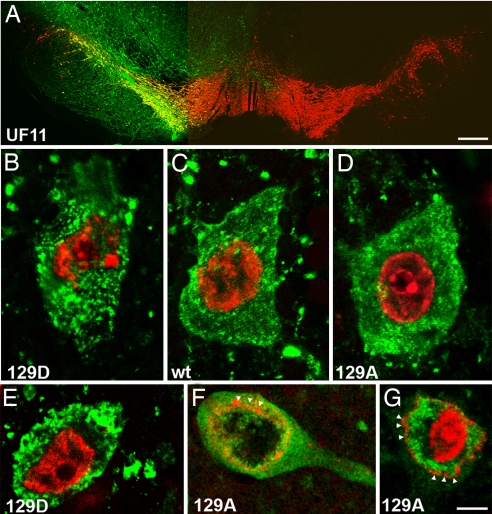
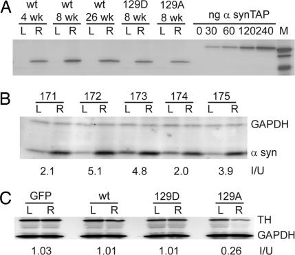
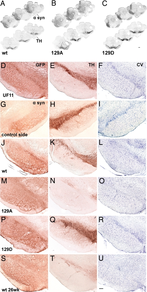
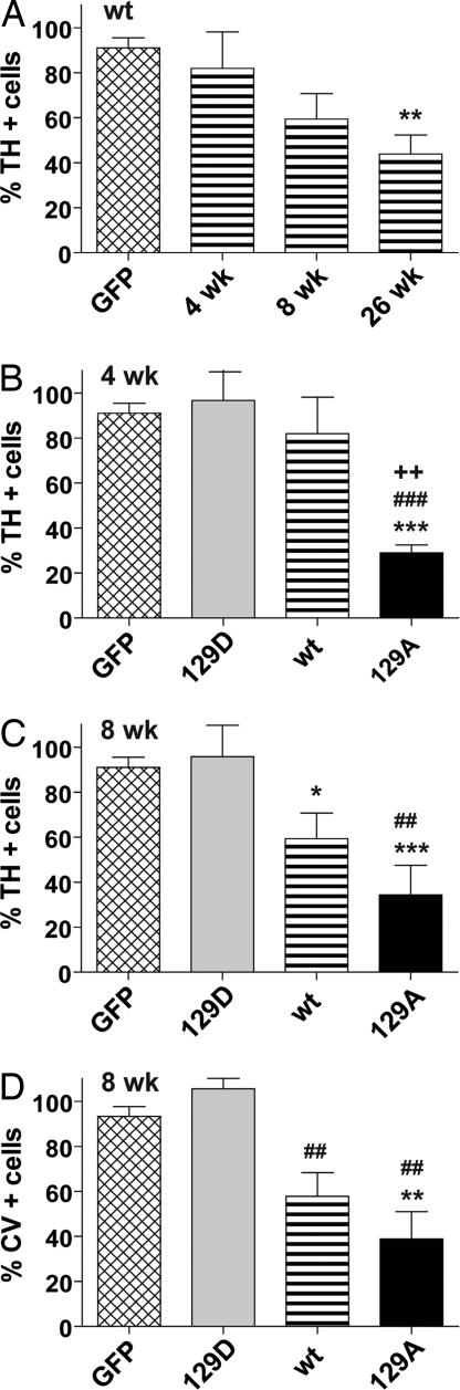
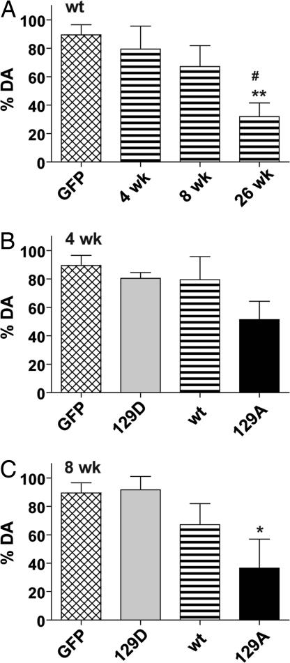
Studies have shown that alpha-synuclein (alpha-syn) deposited in Lewy bodies in brain tissue from patients with Parkinson disease (PD) is extensively phosphorylated at Ser-129. We used recombinant Adeno-associated virus (rAAV) to overexpress human wild-type (wt) alpha-syn and two human alpha-syn mutants with site-directed replacement of Ser-129 to alanine (S129A) or to aspartate (S129D) in the nigrostriatal tract of the rat to investigate the effect of Ser-129 phosphorylation state on dopaminergic neuron pathology. Rats were injected with rAAV2/5 vectors in the substantia nigra pars compacta (SNc) on one side of the brain; the other side remained as a nontransduced control. The level of human wt or mutant alpha-syn expressed on the injected side was about four times the endogenous rat alpha-syn. There was a significant reduction of dopaminergic neurons in the SNc and dopamine (DA) and tyrosine hydroxylase (TH) levels in the striatum of all S129A-treated rats as early as 4 wk postinjection. Nigral DA pathology occurred more slowly in the wt-injected animals, but by 26 wk the wt alpha-syn group lost nigral TH neurons equivalent to the mutated S129A group at 8 wk. In stark contrast, we did not observe any pathological changes in S129D-treated animals. Therefore, the nonphosphorylated form of S129 exacerbates alpha-syn-induced nigral pathology, whereas Ser-129 phosphorylation eliminates alpha-syn-induced nigrostriatal degeneration. This suggests possible new therapeutic targets for Parkinson Disease.


Conflict of interest statement

Conflict of interest statement: N.M. is an inventor of patents related to recombinant AAV technology and owns equity in a gene therapy company that is commercializing AAV for gene therapy applications.

Figures

Fig. 1.
Fig. 1.
Fluorescence microscopy of tyrosine hydroxylase (TH)-positive neurons expressing α-syn mutants. (A) Photomicrographs showing the expression of the GFP transgene on the injected side of rat brain. GFP (green) was expressed in the majority of the TH-positive neurons (red) in SNc and also can be seen in cells of the mesenphalic tegmentum and in the SN pars reticulata. (B–G) Confocal images illustrate α-syn expression in SNc neurons 4 (B–D) and 8 (E–G) wk after injection. In B–G, α-syn is stained green and nuclei are stained with propidium iodide (red). S129D is more prone to make punctate aggregates at 4 wk (B), becoming more dense and larger at 8 wk (E) compared with wt (C) and 129A (D, F, and G). (F and G) Examples of neurodegenerative changes in SNc neurons of 129A-injected animals at 8 wk that have diffuse α-syn immunoreactivity and vacuolated nuclei. containing both diffuse and aggregated α-syn. α-syn was found both inside and outside of the nuclear membrane (arrowheads indicate propidium iodide staining at the nuclear membrane). (Scale bar: A, 0.5 mm; B–G, 5 μm.)
Fig. 2.
Fig. 2.
Measurement of α-syn and TH expression in α-syn-injected animals. (A) Pooled samples of striatal extracts taken from the uninjected left side (L) and injected right side (R) were electrophoresed on acrylamide gels. Fifty micrograms of protein from animals injected with wt, 129A, and 129D at the indicated time points were Western blotted with an antibody specific for human α-syn and a fluorescent secondary antibody, and compared with purified samples of a human α-syn-TAP fusion protein of known concentration. The concentration of α-syn was calculated from the fluorescence measurements by using ImagQuant software. (B) SNc tissue was excised from uninjected (L) and injected (R) sides of individual animals (171–175) that had been injected with wt α-syn at 4 wk postinjection. Fifty micrograms of tissue extract was Western blotted with antibodies that recognize rat α-syn or GAPDH. The ratio of α-syn on the injected and uninjected sides (I/U) was calculated for each animal. (C) Pooled striatal tissue extracts from 4-wk animals was Western blotted with TH and GAPDH antibodies. The amount of TH enzyme was normalized to GAPDH and the ratio of the injected vs. uninjected sides was calculated (I/U). At 4 wk only the 129A samples showed a reduction in TH compared with total protein.
Fig. 3.
Fig. 3.
Photomicrographs showing nigral degeneration in the rAAV-α-syn-injected animals. (A–C) Montages of rostral-to-caudal coronal sections illustrate transduction volume in different experimental groups at 8 wk postinjection (α-syn-stained sections, Upper) and a reduction of TH immunoreactivity on the injected side of parallel sections (Lower). These sections were labeled with standard DAB immunohistochemistry (see Materials and Methods). (D–J) Each row contains neighboring sections, taken from the same animal, that illustrate cells immunostained for GFP (D), α-syn (G, J, M, P, S) and TH (E, H, K, N, Q, T), and CV-stained cells (F, I, L, O, R, U) in the SN at 8 (D–R) and 26 (S–U) wk postinjection. The sections were uninjected (G–I) or injected with UF11 (which expresses GFP, D–F), wt-α-syn (J–L, S–U), 129A (M–O), 129D (P–R). Whereas expression of GFP protein (D–F) or 129D (P–R) did not alter the number of TH-positive neurons or CV-stained neurons, the expression of 129A (M–O) or wt (J–L and S–U) α-syn led to loss of TH-positive neurons and CV staining in the SNc, compared with the contralateral intact side (G–I). (Scale bars: A–D, 1 mm; E–V, 250 μm.)
Fig. 4.
Fig. 4.
Unbiased estimation of nigral TH+ cells in SNc of animals injected with wt or mutant human α-syn. Tissue slices were labeled with antibody to TH (A–C) or stained with cresyl violet (D) as described in Materials and Methods and the percentage of surviving cells was calculated by comparison with the uninjected side in the same animal. The graphs display a comparison of wt injected animals compared with GFP as a function of time postinjection (A), and a comparison of the wt, 129A, and 129D animals compared with GFP at 4 wk (B) and 8 wk (C) postinjection. The GFP control in A–C is the same and includes 4-, 8-, and 26-wk samples as these did not vary significantly. The wt 4-wk and 8-wk groups in B and C are the same as in A and are repeated to facilitate comparison. (D) Eight-week samples stained with cresyl violet. Group ANOVA statistics were for (A) F[3,34] = 5.973, P = 0.002; (B) F[3,32] = 13.53, P = 0.001; (C) F[3, 29] = 9.22, P = 0.0002; (D) F[3,15] = 9.431, P = 0.001. Tukey's post hoc results are indicated as *, **, *** = P < 0.05, 0.01, and 0.001, respectively vs. GFP; #, ##, ### = P < 0.05, 0.01, and 0.001 vs. 129D; ++ = P < 0.01 vs. wt 4 wk.
Fig. 5.
Fig. 5.
Measurement of striatal dopamine. The amount of dopamine in striatal tissue was measured on the injected and uninjected sides of individual animals as described in Materials and Methods and is displayed as the mean percentage of dopamine remaining on the injected side compared with the uninjected side plus standard error. Tukey's post hoc results are indicated as in Fig. 4. Group ANOVA statistics were F[3,28] = 5.949, P = 0.0029 (A); F[3,33] = 2.867, P = 0.0513 (B); and F[3,24] = 4.079, P = 0.0179 (C).

References

    1. Cookson MR. The biochemistry of Parkinson's disease. Annu Rev Biochem. 2005;74:29–52. - PubMed
    1. Eslamboli A, et al. Long-term consequences of human alpha-synuclein overexpression in the primate ventral midbrain. Brain. 2007;130:799–815. - PubMed
    1. Kirik D, et al. Parkinson-like neurodegeneration induced by targeted overexpression of alpha-synuclein in the nigrostriatal system. J Neurosci. 2002;22:2780–2791. - PMC - PubMed
    1. Periquet M, Fulga T, Myllykangas L, Schlossmacher MG, Feany MB. Aggregated alpha-synuclein mediates dopaminergic neurotoxicity in vivo. J Neurosci. 2007;27:3338–3346. - PMC - PubMed
    1. Fredenburg RA, et al. The impact of the E46K mutation on the properties of alpha-synuclein in its monomeric and oligomeric states. Biochemistry. 2007;46:7107–7118. - PubMed

Publication types

LinkOut - more resources