. 2008 Jan 15;105(2):781-5.
doi: 10.1073/pnas.0708061105. Epub 2008 Jan 4.

Rapid cell-cycle reentry and cell death after acute inactivation of the retinoblastoma gene product in postnatal cochlear hair cells

Affiliations

Rapid cell-cycle reentry and cell death after acute inactivation of the retinoblastoma gene product in postnatal cochlear hair cells

Thomas Weber et al. Proc Natl Acad Sci U S A. .

Abstract

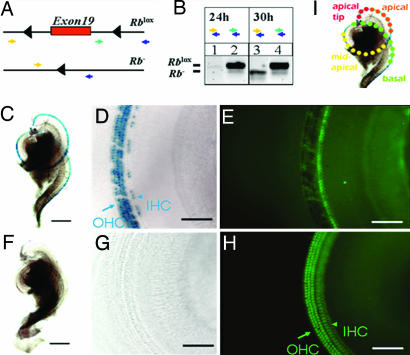
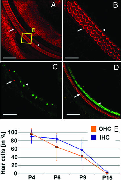
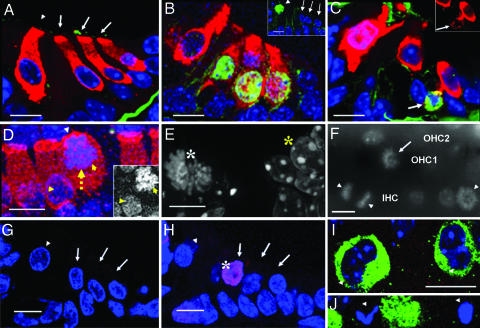
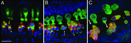
Unlike lower vertebrates, mammals are unable to replace damaged mechanosensory hair cells (HCs) in the cochlea. Recently, ablation of the retinoblastoma protein (Rb) in undifferentiated mouse HC precursors was shown to cause cochlear HC proliferation and the generation of new HCs, raising the hope that inactivation of Rb in postmitotic HCs could trigger cell division and regenerate functional HCs postnatally. Here, we acutely inactivated Rb in nearly all cochlear HCs of newborn mice, using a newly developed HC-specific inducible Cre mouse line. Beginning 48 h after Rb deletion, approximately 40% of HCs were in the S and M phases of the cell cycle, demonstrating an overriding role for Rb in maintaining the quiescent state of postnatal HCs. Unlike Rb-null HC precursors, such HCs failed to undergo cell division and died rapidly. HC clusters were restricted to the less differentiated cochlear regions, consistent with differentiation-dependent roles of Rb. Moreover, outer HCs expressed the maturation marker prestin, suggesting an embryonic time window for Rb-dependent HC specification. We conclude that Rb plays essential and age-dependent roles during HC proliferation and differentiation, and, in contrast to previous hypotheses, cell death after forced cell-cycle reentry presents a major challenge for mammalian HC regeneration from residual postnatal HCs.


Conflict of interest statement

The authors declare no conflict of interest.

Figures

Fig. 1.
Fig. 1.
Inducible recombination of the Rb gene locus. (A) Two targeted Rb alleles. Exon 19 of Rb is flanked by two unrecombined loxP sites (black triangles; Rblox in B Lower). Cre-mediated deletion of exon 19 results in an allele encoding a nonfunctional Rb protein (Rb in B Lower). Two combinations of PCR genotyping primer pairs were used to distinguish the Rblox allele (283-bp band from the green and blue pair) from the Rb allele (260-bp band from the yellow and blue pair). (B) PCR analysis, using cochlear genomic DNA of Atoh1-CreER: Rblox/lox mice given injections on P0. The primer combination used for each lane is indicated. The Rb band was not detected after 24 h (lane 1), but it was detected after 30 h (lane 3). The corresponding Rblox bands from unrecombined cells are shown as controls (lanes 2 and 4). (C–H) Cre activity within the cochlea as visualized by lacZ staining. The mice were given injections on P0 and P1 and analyzed on P6. (C and F) LacZ-positive (blue) staining of cochlear whole mounts from Atoh1-CreER: Rosa26 mice given tamoxifen injections (C) and mock injections (F). (D and G). Higher magnification reveals that Cre-activity was limited to OHCs (arrow) and IHCs (arrowhead) (blue, lacZ staining in D), whereas no lacZ staining was detected in mice given mock injections (G). (E and H) Myosin7a (green) coimmunostaining of regions in D and G reveals the presence of HCs. Note that fluorescence is masked in cells that express lacZ (E compared with H). [Scale bars: C, 0.25 mm (applies to C and F); and D, 80 μm (applies to D, E, G, and H)]. Note that ≈80% of IHCs and ≈90% of OHCs were lacZ-positive when induced at P0 and P1 under these conditions (14). (I) Illustration of HCs and their locations in different regions within a cochlear whole mount from basal (green dots) to apical (red dots). The apical tip contained the least differentiated HCs at the times analyzed in this study (P4–P6 and P9), whereas the basal HCs were more mature. Consistently, in subsequent figures (unless stated otherwise), arrows denote OHCs, and arrowheads denote IHCs.
Fig. 2.
Fig. 2.
HC loss in Rb−/− mice. (A) Confocal z projections reveal the normal morphology of an Rb−/− cochlear whole mount on P3 after tamoxifen injection on P0 and P1. [Scale bar, 100 μm (refers to A, C and D)]. Phalloidin staining (red). Arrows indicate OHCs; arrowheads indicate IHCs. (B) The region in the yellow box in A at a higher magnification allows the visualization of the orderly stereocilia (V-shapes) of OHCs (three rows) and IHCs (one row). (Scale bar, 20 μm.) (C) On P15 Rb−/− mice display a severe loss of IHCs and OHCs. Prestin (red) and myosin7a (green). (D) Rb+/− littermates exhibit no cochlear abnormalities on P15. (E) Progressive loss of OHCs (blue) and IHCs (orange) from P4 to P15 in Rb−/− mice. Surviving HCs in Rb−/− mice were normalized to those in Rb+/− littermates at corresponding ages and are depicted as a percentage of Rb−/−/Rb+/−. Curves were fitted by using linear regression of mean values. Error bars indicate standard deviations. The number of cochlear regions in at least three mice analyzed and statistical significance (Rb−/− vs. Rb+/−, Student's t test) are as follows: on P4, IHCs, n = 7 (P > 0.05), and OHCs, n = 8 (P > 0.05); on P6, IHCs, n = 7 (P < 0.01), and OHCs, n = 5 (P < 0.05); on P9, IHCs, n = 12 (P < 0.01), and OHCs, n = 12 (P < 0.01); and on P15, IHCs, n = 6 (P < 0.01), and OHCs, n = 6 (P < 0.01).
Fig. 3.
Fig. 3.
Rb−/− HCs in the S and M phases of the cell cycle and undergoing cell death. (A–C) Confocal images of sections through the organ of Corti, myosin7a immunostaining marks HCs (red, myosin7a; blue, DAPI). (A and B) Overlay in a BrdU pulse-labeling experiment on P4 (green, BrdU). (A) No BrdU staining in HC nuclei of Rb+/− mice (none of 597 HCs, n = 3). Basilar membrane staining is background. Arrows indicate OHCs; the arrowhead indicates IHC. (B) Strong BrdU staining in three OHC nuclei of Rb−/− mice. A total of 261 of 659 HCs (39.6%) and no SC nuclei were BrdU-positive (n = 2). Staining of myosin7a in the nucleus of the OHC at the right might indicate disruption of the nuclear envelope and the beginning of cell death. (Inset) BrdU labeling of an IHC (green, arrowhead) of a basal turn cochlear section at P4; this cell is presumed IHC because of its expression of myosin7a (data not shown) and its position. (C) Phosphorylated histone H3 labeling (green, arrow) of Rb−/− OHCs on P5. (Inset) myosin7a staining (red channel only) of the same cell (the arrow indicates positive for phosphorylated histone H3), demonstrating that it is an HC. (D) Confocal whole-mount z projection of Rb−/− IHCs. Blue, DAPI; red, myosin7a. The yellow arrow indicates nuclear migration from the basal to the apical pole of the cell. Yellow arrowheads indicate migrating and normal nuclei of IHCs. The white arrowhead indicates the intact cuticular plate at the apical pole. (Inset) DAPI channel (in gray) of the same IHC nuclei marked with yellow arrowheads. (E) High-resolution deconvoluted fluorescence image of a basal Rb−/− HC nucleus on P5 stained with DAPI (white asterisk); individual chromosomes in metaphase can be seen. Adjacent interphase nuclei are shown for comparison (yellow asterisk). (F) DAPI (gray) fluorescence image (omitting myosin7a signal) of an Rb−/− cochlear whole mount from an apical turn on P5 displaying mitotic IHCs (arrowheads) and OHCs (arrows). Note an IHC in anaphase (two opposing arrowheads). Rows of OHC1, OHC2, and IHC are labeled based on DAPI and myosin7a staining (data not shown). (G and H) Confocal images of TUNEL-labeled (red, asterisk) cross-sections through the organ of Corti on P4. (G) Rb+/−. (H) Rb−/−. Blue, DAPI. Arrows and arrowheads indicate OHCs and IHCs, respectively. TUNEL-positive HCs were detected at presumed locations of IHCs (data not shown). (I and J) Confocal z projection of an Rb−/− apical cochlear whole mount on P6. Blue, DAPI; green, myosin7a. Arrowheads indicate kidney-shaped and abnormal, partially fragmented nuclei of HCs. (Scale bars: 10 μm.)
Fig. 4.
Fig. 4.
HC clusters in the most apical cochlear turns and prestin expression. Confocal z projection of an Rb−/− cochlear whole mount on P9. Green, myosin7a; red, prestin; blue, DAPI. (A) Basal cochlear turn with significant HC loss. The dotted red arrow indicates a mitotic OHC that has rounded up, displaying nuclear migration. the yellow arrow and arrowhead indicate apical and basal poles, respectively, of an apparently normal OHC with the mature prestin expression pattern, which is reduced or absent at the apical and basal poles. The red arrow indicates the abnormal subcellular distribution of the membrane protein prestin in the same cochlear region. (B) Mid-apical cochlear turn. The white arrow indicates a rounded-up OHC next to apparently normal OHCs; the yellow arrowhead and arrow are as in A. Note that apical HCs are larger and display a more distinct prestin-free basal pole beginning at the supranuclear region. (C) Most apical cochlear tip. Note HC loss and OHC clusters (white arrows) with abnormal prestin expression. (Scale bars: 25 μm.)

References

    1. Lee YS, Liu F, Segil N. Development (Cambridge, UK) 2006;133:2817–2826. - PubMed
    1. Mantela J, Jiang Z, Ylikoski J, Fritzsch B, Zacksenhaus E, Pirvola U. Development (Cambridge, UK) 2005;132:2377–2388. - PMC - PubMed
    1. Ruben RJ. Acta Otolaryngol. 1967;220(Suppl):1–44. - PubMed
    1. Kelley MW. Brain Res. 2006;1091:172–185. - PubMed
    1. Corwin JT, Oberholtzer JC. Neuron. 1997;19:951–954. - PubMed

Publication types

LinkOut - more resources