. 2008 Apr 1;94(7):2639-53.
doi: 10.1529/biophysj.107.123257. Epub 2008 Jan 4.

The actions of calcium on hair bundle mechanics in mammalian cochlear hair cells

Affiliations

The actions of calcium on hair bundle mechanics in mammalian cochlear hair cells

Maryline Beurg et al. Biophys J. .

Abstract

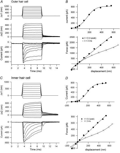
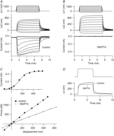
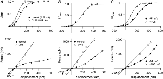
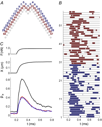
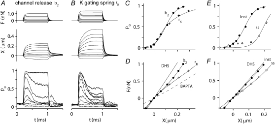
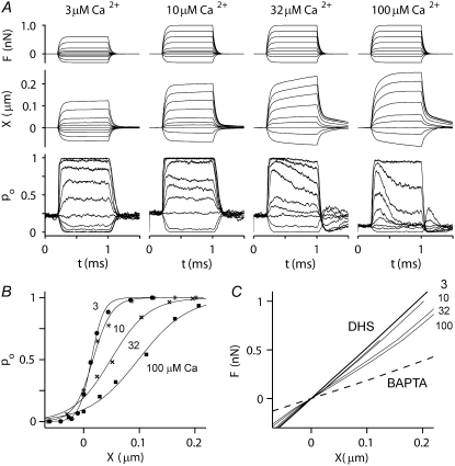
Sound stimuli excite cochlear hair cells by vibration of each hair bundle, which opens mechanotransducer (MT) channels. We have measured hair-bundle mechanics in isolated rat cochleas by stimulation with flexible glass fibers and simultaneous recording of the MT current. Both inner and outer hair-cell bundles exhibited force-displacement relationships with a nonlinearity that reflects a time-dependent reduction in stiffness. The nonlinearity was abolished, and hair-bundle stiffness increased, by maneuvers that diminished calcium influx through the MT channels: lowering extracellular calcium, blocking the MT current with dihydrostreptomycin, or depolarizing to positive potentials. To simulate the effects of Ca(2+), we constructed a finite-element model of the outer hair cell bundle that incorporates the gating-spring hypothesis for MT channel activation. Four calcium ions were assumed to bind to the MT channel, making it harder to open, and, in addition, Ca(2+) was posited to cause either a channel release or a decrease in the gating-spring stiffness. Both mechanisms produced Ca(2+) effects on adaptation and bundle mechanics comparable to those measured experimentally. We suggest that fast adaptation and force generation by the hair bundle may stem from the action of Ca(2+) on the channel complex and do not necessarily require the direct involvement of a myosin motor. The significance of these results for cochlear transduction and amplification are discussed.


Figures

FIGURE 1
FIGURE 1
Method of mechanical stimulation, showing four rows of hair bundles, one IHC at the bottom and three OHCs with the flexible fiber connected to a first-row OHC bundle by a Sylgard bead. The fiber is attached to a piezoelectric actuator and is introduced along the long axis of the cochlea, here from the right. P8 rat.
FIGURE 2
FIGURE 2
Model of an OHC bundle. (A) Geometry of a simulated hair bundle that contains three rows, with 29 stereocilia per row. The tallest stereocilia are nearly upright, whereas the shorter stereocilia are tilted toward the taller stereocilia. (B) There are three structural components: stereocilia, tip link (assembly), and horizontal connector. The channels are located at the upper end of each tip link. (C) The kinetic scheme used in the model for the MT channel has five closed (C) and five open (O) states, with the rate constants between C and O dependent on the mechanical stimulus and the transitions between each of the C (or O) states dependent on binding of one to four calcium ions. As Ca2+ binds to the channel, it requires an additional force, fCa, to open a channel, and the gating spring is released by the fixed amount, b2.
FIGURE 3
FIGURE 3
Hair-bundle displacements and MT currents in OHCs and IHCs. (A) OHC motion of the fixed end of the fiber cemented to the piezoelectric actuator (Δx1, upper), the end attached to an OHC bundle (Δx2, middle), and MT current (lower). The oscillations in the displacement and current records are attributable to slight underdamping of the stimulator. (B) MT current-displacement relationship (upper), with peak current plotted against instantaneous displacement measured at the peak of the current, and force-displacement relationships (lower) from records in A. The force is plotted against displacements measured 0.5 ms (instantaneous) and 3 ms (steady state) after stimulus onset. KHB = 5 mN·m−1, P7 rat. (C) IHC motion of the fixed end of a fiber (Δx1, upper), the end attached to the inner hair cell bundle (Δx2, middle) and MT current (lower). (D) MT current-displacement relationship (upper), with the peak current plotted against instantaneous displacement at the peak current, and force-displacement relationships (lower) from records in C. The force is plotted against displacements measured 0.6 ms (instantaneous) and 3 ms (steady state) after stimulus onset. KHB = 1.5 mN·m−1, P11 rat. Stiffness of flexible fiber: 1.6 mN·m−1 (A); 2.3 mN·m−1 (C).
FIGURE 4
FIGURE 4
Effects of BAPTA on hair-bundle stiffness of an OHC. (A) Control motion of the fixed end of the fiber (Δx1, upper), the end attached to the hair bundle (Δx2, middle), and MT current (lower). (B) Same measurements after a 2-s puff of a BAPTA-containing saline that abolished the MT current. (C) MT current plotted against instantaneous displacement (upper) and force plotted against steady-state displacement relationships (lower). The force-displacement plots are given before (solid circles, KHB = 2.5 mN·m−1) and after BAPTA application (crosses, KHB = 1.1 mN·m−1). P8 rat, IMAX = 490 pA. (D) Comparison of the two displacements of similar size shows the extra secondary component before BAPTA treatment. Stiffness of fiber, 1.9 mN·m−1.
FIGURE 5
FIGURE 5
Effects of extracellular calcium on the hair-bundle mechanics in an OHC. (A) Motion of the fixed end of the fiber cemented to the piezoelectric actuator (Δx1, upper), the free end of the fiber attached to the hair bundle (Δx2, middle), and MT current (lower) in 1.5 mM extracellular calcium. (B) Motion of the fixed end of the fiber (Δx1, upper), the end attached to the hair bundle (Δx2, middle), and MT current (lower) in 0.02 mM extracellular calcium. (C, upper) Current-displacement relationships in 1.5 mM calcium (solid circles, prior control; open circles, wash) and in 0.02 mM calcium (crosses). (C, lower) Force-displacement relationships, with symbols the same as in upper. Fast-adaptation time constants (dashed lines): 0.44 ms for 1.5 mM calcium; 1.7 ms for 0.02 mM calcium. Secondary creep in displacement (dashed lines): 0.43 ms, 1.5 mM calcium; 0.70 ms; 0.02 mM calcium. P10 rat, IMAX = 800 pA, KHB = 8 mN·m−1 in 1.5 mM calcium. Stiffness of fiber, 2.4 mN·m−1.
FIGURE 6
FIGURE 6
Effects of DHS and depolarization on the hair-bundle mechanics. (A, upper) IHC, with MT current scaled to its maximum value, IMAX, plotted against displacement in control saline (solid circles, prior control) and in saline containing 0.2 mM DHS (crosses). (A, lower) Force-displacement relationships, with symbols the same as in A, upper. The current-displacement relationships have been fitted with single Boltzmann functions (see Methods), with IMAX = 655 pA (control) and 40 pA (DHS). Note that the DHS plot is shifted negative, which is consistent with a reduction in resting intracellular calcium due to channel block. KHB = 3.4 mN·m−1 in control and 4.9 mN·m−1 in DHS. P11 rat. (B, upper) OHC, with MT current scaled to its maximum value, IMAX, plotted against displacement in control saline. IMAX = 430 pA. (B, lower) Force-displacement relationships in control (solid circles) and in 0.2 mM DHS (crosses). The MT current in DHS was too small to be plotted. KHB = 4.2 mN·m−1 in control and 14.4 mN·m−1 in DHS. P11 rat. (C, upper) OHC, with MT current scaled to its maximum value, IMAX, plotted against displacement at −84 mV holding potential (solid circles) and at +106 mV (crosses). (C, lower) Force-displacement relationships with symbols the same as in C, upper. IMAX and KHB = 560 pA and 1.7 mN·m−1, respectively, at −84 mV, and 610 pA and 3.6 mN·m−1, respectively, at +106 mV. P11 rat. Stiffness of flexible fiber for all three cells, 1.6 mN·m−1.
FIGURE 7
FIGURE 7
Simulated responses of an OHC. (A) Top view of stereocilia showing numbering of MT channels in the first row (blue, 1–29) and second row (red, 30–58). MT channels are located solely at the top of each tip link, so the third row of stereocilia has no channels. (B) The channel numbers correspond to the numbers on the ordinate in the plot, showing the activity of each individual channel during a single force step of 0.9 nN, which was applied at t = 0.2 ms. The force was distributed equally to the tips of the tallest stereocilia. (C) Time course of the force step, F (upper curve), displacement, X (middle curve), and average probability of opening, pO (lower), computed from the individual responses in B. The average (pO) is identical for the first row (blue) and second row (red), the sum of the two responses shown in black.
FIGURE 8
FIGURE 8
Simulated MT currents and hair-bundle mechanics on two different models. (A) Channel release model (b2): applied step force (F), bundle tip displacement (X), and the probability of MT channel opening (pO). The X and pO responses are averages of 10 simulations. Intracellular calcium at the fast-adaptation site with the MT channel open was 100 μM. (B) Calcium-induced reduction in the gating spring (rK): same simulations as in A. rK = 0.8 represents a maximal reduction in stiffness of 80%. (C) pO-X relationships of the channel on the “channel release” model (b2, solid circles) and on the “reduction in gating spring stiffness” model (rK, crosses). Results fitted with a single Boltzmann. (D) Force-displacement (F-X) relationships of the bundle for the two models. In addition, two passive conditions with no channel activity were also modeled. To simulate a hair cell treated with DHS, all the MT channels were blocked. To simulate a BAPTA-treated hair cell, the tip links were removed. KHB at X = 0 is 3.6 mN·m−1 (control; b2), 3.9 mN·m−1 (control; rk), 5.3 mN·m−1 (DHS), and 1.8 mN·m−1 (BAPTA). (E) pO-X relationships on the channel release model at the peak of the response (solid circles, inst) and after 1 ms (open circles, ss). (F) F-X relationships on the channel release model at the peak of the response (solid circles, inst) and after 1 ms (open circles, ss). Note that the instantaneous plot is close to that without channel activity in the presence of the blocker DHS.
FIGURE 9
FIGURE 9
Simulated effect of calcium on MT channel and hair-bundle mechanics. (A) Probability of MT channel opening, pO (lower) and hair-bundle displacements, X (middle), for force steps (upper) at four different calcium concentrations: 3, 10, 32, and 100 μM at the fast-adaptation site (CFA) with the MT channel open. These calcium concentrations are approximately equivalent to 20 μM to 1.2 mM externally. Note the change in the speed of adaptation and of the secondary component of bundle displacement as the calcium increases. The adaptation time constants for small changes in pO for different values of CFA were 0.12 ms at 100 μM; 0.25 ms at 30 μM; 0.6 ms at 10 μM; and >1 ms at 3 μM calcium. (B) Effects of calcium on pO-X relationships for the four calcium concentrations. All relationships were fitted with a first-order Boltzmann equation. (Left to right) 3 μM (solid circles), 10 μM (asterisks), 32 μM (crosses), and 100 μM (solid squares) intracellular calcium. Note that the 10–90% operating range increased from 47 to 200 nm as CFA was increased from 3 to 100 μM. (C) F-X relationships for four different calcium concentrations (3, 10, 32, and 100 μM). Black lines are from the passive hair bundle with DHS (solid line) and BAPTA treatment (dashed line). With progressively increasing calcium concentration, the F-X relationship deviates further from the DHS curve. Simulations in this figure were performed only with the b2 model.
FIGURE 10
FIGURE 10
Theoretical contribution of the myosin motor on the channel release model. (A) Rebound of open probability after a negative step (pO) as a function of the duration of the step in the presence (black lines) and absence (red lines) of the myosin motor. The envelope of the rebound is an indicator of the adaptation process, which is little affected by removal of the myosin. (B) Effects of a positive 0.5-nN adapting step on the response onsets in the presence (black) and absence (red) of myosin. Note that lack of myosin does not affect the fast-adaptation time constant for small stimuli. The bundle displacement is 140 nm. (C) pO-X relationships under control conditions (solid circles) and after the 0.5-nN adapting step (open circles) in the presence (black) and absence (red) of myosin. The shift in the pO-X relationship is 110 nm. In these simulations, the rate constant for myosin-driven adaptation, kA, was 0.05 nm·ms−1·pN−1.
FIGURE 11
FIGURE 11
Kinetic contribution of myosin to MT channel responses (A) Simulated probability of MT channel opening, pO (lower) and hair-bundle displacements, X (middle), for a force step (upper) for CFA = 3 μM calcium shown on a longer time scale. The size of the force step was chosen to evoke pO ≈ 0.5. Note the slow changes in pO and X that are fit with a time constant of 12 ms. No fast component attributable to channel adaptation was visible. (B) Simulated probability of MT channel opening, pO (lower) and hair-bundle displacements, X (middle), for a force step (upper) at CFA = 10 μM. In addition to the slow component of adaptation, the change in pO now had a fast component of time constant 0.6 ms due to the effects of calcium on the MT channel. In these simulations, the rate constant for myosin-driven adaptation, kA, was 0.05 nm·ms−1·pN−1. (C) Experimental response of an OHC to an extended displacement step. The adaptation was fitted with two components with time constants of 0.3 ms and 12 ms. P8 rat, 1.5 mM external calcium.

References

    1. Pickles, J. O., S. D. Comis, and M. P. Osborne. 1984. Cross-links between stereocilia in the guinea pig organ of Corti, and their possible relation to sensory transduction. Hear. Res. 15:103–112. - PubMed
    1. Hudspeth, A. J. 1989. How the ear's works work. Nature. 341:397–404. - PubMed
    1. Ohmori, H. 1985. Mechano-electrical transduction currents in isolated vestibular hair cells of the chick. J. Physiol. 359:189–217. - PMC - PubMed
    1. Beurg, M., M. G. Evans, C. M. Hackney, and R. Fettiplace. 2006. A large-conductance calcium-selective mechanotransducer channel in mammalian cochlear hair cells. J. Neurosci. 26:10992–11000. - PMC - PubMed
    1. Eatock, R. A. 2000. Adaptation in hair cells. Annu. Rev. Neurosci. 23:285–314. - PubMed

Publication types

MeSH terms

LinkOut - more resources