

Crystallographic study of hydration of an internal cavity in engineered proteins with buried polar or ionizable groups
- PMID: 18178652
- PMCID: PMC2275713
- DOI: 10.1529/biophysj.107.122473
Crystallographic study of hydration of an internal cavity in engineered proteins with buried polar or ionizable groups
Abstract
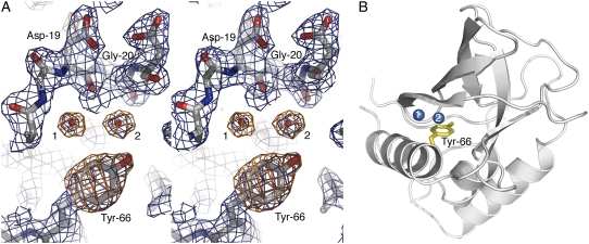
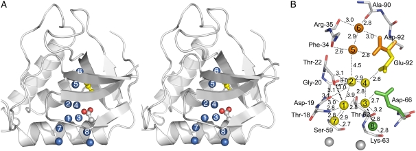
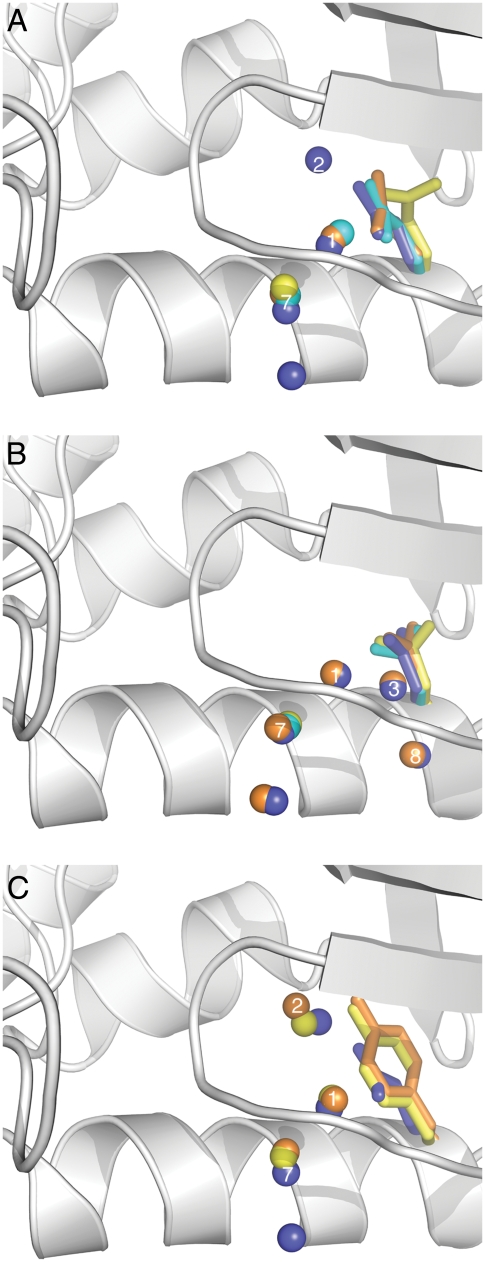
Although internal water molecules are essential for the structure and function of many proteins, the structural and physical factors that govern internal hydration are poorly understood. We have examined the molecular determinants of internal hydration systematically, by solving the crystal structures of variants of staphylococcal nuclease with Gln-66, Asn-66, and Tyr-66 at cryo (100 K) and room (298 K) temperatures, and comparing them with existing cryo and room temperature structures of variants with Glu-66, Asp-66, Lys-66, Glu-92 or Lys-92 obtained under conditions of pH where the internal ionizable groups are in the neutral state. At cryogenic temperatures the polar moieties of all these internal side chains are hydrated except in the cases of Lys-66 and Lys-92. At room temperature the internal water molecules were observed only in variants with Glu-66 and Tyr-66; water molecules in the other variants are probably present but they are disordered and therefore undetectable crystallographically. Each internal water molecule establishes between 3 and 5 hydrogen bonds with the protein or with other internal water molecules. The strength of interactions between internal polar side chains and water molecules seems to decrease from carboxylic acids to amides to amines. Low temperature, low cavity volume, and the presence of oxygen atoms in the cavity increase the positional stability of internal water molecules. This set of structures and the physical insight they contribute into internal hydration will be useful for the development and benchmarking of computational methods for artificial hydration of pockets, cavities, and active sites in proteins.
Figures
References
-
- Park, S., and J. G. Saven. 2005. Statistical and molecular dynamics studies of buried waters in globular proteins. Proteins Struct. Funct. Genet. 60:450–463. - PubMed
-
- Rose, G. D., W. B. Young, and L. M. Gierasch. 1983. Interior turns in globular proteins. Nature. 304:655–657. - PubMed
-
- Warshel, A. 1998. Electrostatic origin of the catalytic power of enzymes and the role of preorganized active sites. J. Biol. Chem. 273:27035–27038. - PubMed
Publication types
MeSH terms
Substances
Grants and funding
LinkOut - more resources
Full Text Sources
Other Literature Sources
