. 2008 Mar;190(6):2172-82.
doi: 10.1128/JB.01657-07. Epub 2008 Jan 4.

Genomic characterization of mycobacteriophage Giles: evidence for phage acquisition of host DNA by illegitimate recombination

Affiliations

Genomic characterization of mycobacteriophage Giles: evidence for phage acquisition of host DNA by illegitimate recombination

Peter Morris et al. J Bacteriol. 2008 Mar.

Abstract

A characteristic feature of bacteriophage genomes is that they are architecturally mosaic, with each individual genome representing a unique assemblage of individual exchangeable modules. Plausible mechanisms for generating mosaicism include homologous recombination at shared boundary sequences of module junctions, illegitimate recombination in a non-sequence-directed process, and site-specific recombination. Analysis of the novel mycobacteriophage Giles genome not only extends our current perspective on bacteriophage genetic diversity, with more than 60% of the genes unrelated to other mycobacteriophages, but offers novel insights into how mosaic genomes are created. In one example, the integration/excision cassette is atypically situated within the structural gene operon and could have moved there either by illegitimate recombination or more plausibly via integrase-mediated site-specific recombination. In a second example, a DNA segment has been recently acquired from the host bacterial chromosome by illegitimate recombination, providing further evidence that phage genomic mosaicism is generated by nontargeted recombination processes.


Figures
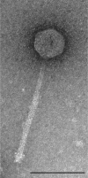
FIG. 1.
FIG. 1.
Morphology of mycobacteriophage Giles virions. Shown is an electron micrograph of mycobacteriophage Giles particles negatively stained with uranyl acetate. Scale bar = 100 nm.
FIG. 2.
FIG. 2.
Organization of the mycobacteriophage Giles genome. The Giles genome is represented by horizontal lines, with putative genes shown as boxes above (transcribed rightward) or below (transcribed leftward); the number of each gene is shown within its box. All genes have been sorted into phamilies (Phams) of related sequences using the computer program “Phamerator” (S. Cresawn, R. W. Hendrix, and G. F. Hatfull, unpublished data); the phamily number is displayed above each gene, and the boxes are color coordinated accordingly. Note that the Pham numbers differ from those described previously (12). Putative gene functions are noted. The positions of putative transcription promoters as identified by DNA Master (http://cobamide2.bio.pitt.edu) are shown as arrows.
FIG. 3.
FIG. 3.
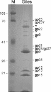
Virion proteins of mycobacteriophage Giles. Purified Giles particles were denatured, and the proteins were separated by SDS-PAGE. Markers (M) are designated by their masses in kDa. Putative assignments of bands to Giles gene products were determined by mass spectrometry and N-terminal sequence analysis of individual bands.
FIG. 4.
FIG. 4.
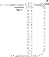
Predicted pseudoknot at the end of the gene 16 mRNA. Bioinformatic analysis suggests that there is a programmed +1 translational frameshift near the end of gene 16 that would shift ribosomes translating gene 16 into the gene 17 reading frame (see the text). We found the illustrated potential pseudoknot and Shine-Dalgarno-like (“S-D”) sequence in the immediate vicinity of the end of gene 16.
FIG. 5.
FIG. 5.
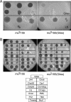
Analysis of Giles lysogens of M. smegmatis. (A) Top agar lawns were prepared with either a putative Giles lysogen or nonlysogenic mc2155 (as indicated), and 5 μl of serial dilutions of phage L5 or Giles was spotted onto the lawn. Giles lysogens are immune to superinfection by Giles, and killing is only observed at the highest phage dose (corresponding to 1010 PFU). (B) A Giles lysogen was similarly tested for immunity to a collection of other mycobacteriophages, all of which infect the lysogen and nonlysogen similarly. A key to the phages tested is shown below.
FIG. 6.
FIG. 6.
Positioning of integration functions in the Giles structural operon. The Giles integration functions are atypically positioned among structural protein genes and the lysis genes. Shared Giles and Halo genes are joined by red shading, and known Giles virion proteins are labeled. While Halo and Giles are not globally related, the alignment illustrates the typical location of the integration cassette at the end of the structural gene operon in Halo, with the lysis and structural genes all to the left of int. In Giles, the integration cassette has moved into an atypical position within the structural operon, with the lysis genes to its right. Pham designations are shown above the genes as described in the legend to Fig. 2.
FIG. 7.
FIG. 7.
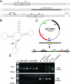
Integration functions of mycobacteriophage Giles. (A) Nucleotide sequence of the intergenic region between Giles genes 28 and 29 (coordinates 24990 to 25285) that includes the attP site (Fig. 2). The 46-bp common core shared with the M. smegmatis genome is shown as a thick horizontal line with the single base difference boxed; the coordinates of the common core in Giles are 25134 to 25179; putative arm-type integrase binding sites (P1 to P4) are also shown. Located between the common core and the right arm-type binding sites is a putative stem-loop terminator for rightward transcription. (B) Structure of M. smegmatis tRNAPro (Msmeg_3734), which includes the Giles attB site with an arrow indicating the position corresponding to the 5′ position of the common core. The tRNA position that changes following Giles integration as a consequence of the base difference is circled. (C) Organization of integration-proficient plasmid pGH1000A and its integration into the M. smegmatis chromosome. (D) PCR amplification of attachment junctions of Giles lysogen and integrated plasmids. Isolated colonies from transformations with Giles integrating vectors and control plasmids, both integrating and extrachromosomal, and a single colony from the Giles lysogen were resuspended in 200 μl sterile distilled H2O and boiled at 95°C for 6 min, and 1 μl was used as a template in PCRs with primer pairs that amplify either a 770-bp attL fragment or an 842-bp attB fragment. The attL product is amplified from the Giles lysogen and from all Giles transformants; the attB product is obtained with control colonies and wild-type mc2155.
FIG. 8.
FIG. 8.
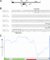
Acquisition of host DNA by illegitimate recombination. (A) Giles gene 79 encodes a putative 103-residue protein with sequence similarity to M. smegmatis MetE; residues 20 to 86 of Giles gp79 are identical to residues 493 to 559 of MetE. (B) Nucleotide sequence comparison showed that the central part of Giles gene 79 contains a 203-bp sequence block that is 100% identical to a central part of the M. smegmatis metE gene (Msmeg_6638). It is not known if Giles gp79 is functional, but the common DNA sequence has apparently been acquired from a mycobacterial chromosome (possibly M. smegmatis) by illegitimate recombination. (C) A transition in GC% at the right end of Giles gene 79 likely reflects an illegitimate-recombination event at the right end of the shared nucleotide block. The GC%s of both the M. smegmatis and Giles genomes are 63 to 65%, but the right end of the Giles genome has a substantially lower GC%. The panel corresponds to approximately 1.5 kbp at the extreme right end of the Giles genome. The reduction in GC% occurs at coordinate ∼54000, and the GC% of the segment between there and the end of the genome is about 50%.

References

    1. Angly, F. E., B. Felts, M. Breitbart, P. Salamon, R. A. Edwards, C. Carlson, A. M. Chan, M. Haynes, S. Kelley, H. Liu, J. M. Mahaffy, J. E. Mueller, J. Nulton, R. Olson, R. Parsons, S. Rayhawk, C. A. Suttle, and F. Rohwer. 2006. The marine viromes of four oceanic regions. PLoS Biol. 4e368. - PMC - PubMed
    1. Baranov, P. V., R. F. Gesteland, and J. F. Atkins. 2002. Recoding: translational bifurcations in gene expression. Gene 286187-201. - PubMed
    1. Brussow, H. 2001. Phages of dairy bacteria. Annu. Rev. Microbiol. 55283-303. - PubMed
    1. Campbell, A. 1958. The different kinds of transducing particles in the lambda-gal system. Cold Spring Harbor Symp. Quant. Biol. 2383-84. - PubMed
    1. Casas, V., and F. Rohwer. 2007. Phage metagenomics. Methods Enzymol. 421259-268. - PubMed

Publication types

MeSH terms

Associated data

LinkOut - more resources