. 2008 Mar;7(3):471-82.
doi: 10.1128/EC.00409-07. Epub 2008 Jan 4.

A class II histone deacetylase acts on newly synthesized histones in Tetrahymena

Affiliations

A class II histone deacetylase acts on newly synthesized histones in Tetrahymena

Joshua J Smith et al. Eukaryot Cell. 2008 Mar.

Abstract

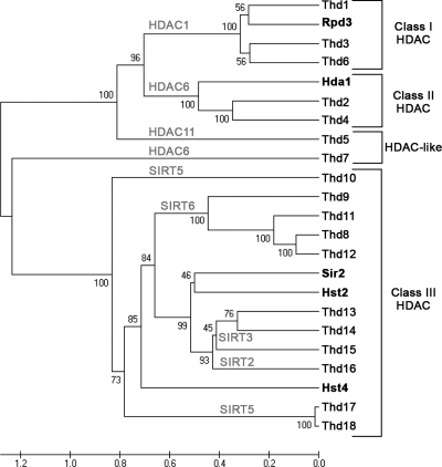
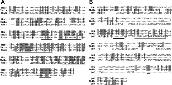
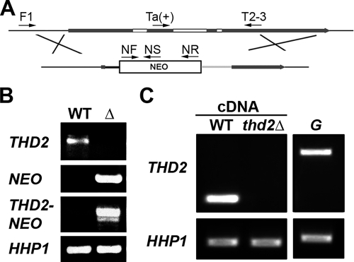
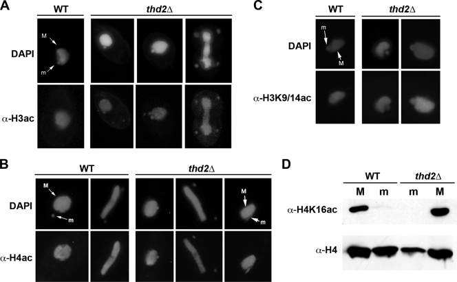
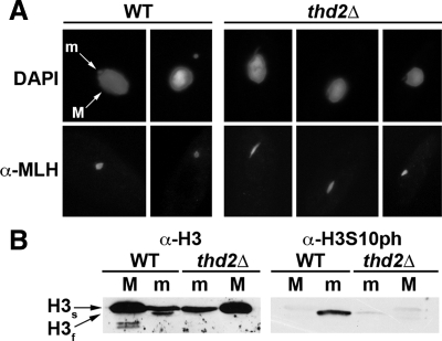
Newly synthesized histones are acetylated prior to their deposition into nucleosomes. Following nucleosome formation and positioning, they are rapidly deacetylated, an event that coincides with further maturation of the chromatin fiber. The histone deacetylases (HDACs) used for histone deposition and de novo chromatin formation are poorly understood. In the ciliate Tetrahymena thermophila, transcription-related deacetylation in the macronucleus is physically separated from deposition-related deacetylation in the micronucleus. This feature was utilized to identify an HDAC named Thd2, a class II HDAC that acts on newly synthesized histones to remove deposition-related acetyl moieties. The THD2 transcript is alternatively spliced, and the major form contains a putative inositol polyphosphate kinase (IPK) domain similar to Ipk2, an enzyme that promotes chromatin remodeling by SWI/SNF remodeling complexes. Cells lacking Thd2, which retain deposition-related acetyl moieties on new histones, exhibit chromatin and cytological phenotypes indicative of a role for Thd2 in chromatin maturation, including the proteolytic processing of histone H3.


Figures

FIG. 1.
FIG. 1.
Phylogenetic tree of the putative Thds (unweighted-pair group method using average linkages). Shown is an alignment of the putative HDAC domains of the 18 Thd protein sequences. S. cerevisiae Rpd3 (class I), Hda1 (class II), Sir2, Hst2, and Hst4 (class III) were used as references to sort the putative Thd proteins into their respective classes (boldface). The positions of human homologs are shown in light-gray capital letters. (Note: HDAC6 and SIRT5 appear twice due to construction of the tree with yeast HDAC domains that are less similar to Thd proteins than those of humans.)
FIG. 2.
FIG. 2.
GFP-Thd2 is localized to the micronucleus. Live-cell images of Tetrahymena transformed with a GFP-Thd2 fusion construct and viewed using the fluorescein isothiocyanate channel to detect GFP signal. The positions of nuclei in each cell were visualized by staining them with DAPI. GFP expressed alone, without fusion to another protein, remained in the cytoplasm; the regions devoid of fluorescence correspond to the macronucleus (M) and the micronucleus (m). Cells expressing GFP-Thd2 showed fluorescence in both the macronucleus and micronucleus, which persisted throughout the cell cycle (see the mitotically dividing cell in the right panel).
FIG. 3.
FIG. 3.
Thd2a is a splice variant of THD2 that contains both an HDAC and an IPK domain. (A) Diagram of the THD2 locus with the flanking sequence (thin black), coding sequence (thick gray) with exons labeled below (E1 to E4), and introns (white) labeled below (i1 to i3). The arrows represent primers used to amplify splice variants of THD2. Displayed below the THD2 sequence is a schematic of the mRNA splice variants for Thd2a (only exons E1, E2, and E4) and Thd2b. The locations of putative HDAC (light gray) and putative IPK (dark gray) domains are indicated. (B) Protein sequence alignment of Thd2a and Thd2b. HDAC domain, light gray; IPK domain, dark gray; change in coding sequence due to splicing frameshift, #; and stop codon, *. (C) RT-PCR of wild-type vegetatively growing (V) and starved (S) cells. PCR on a cDNA template using primers Ta(+) and T2(−) was used to detect THD2a transcripts, and primers Tb(+) and T2(−) were used to detect the THD2b variant transcripts. The cDNA was made with (+) and without (−) RT as a control for genomic DNA contamination. Genomic DNA (G) was amplified as a control. RT-PCR yielded two bands for THD2b; the fastest-migrating band corresponds to THD2b (arrow). (Note: THD2b amplification was detected only with an additional seven cycles of PCR amplification over that used for THD2a detection.)
FIG. 4.
FIG. 4.
Thd2 is a class II HDAC with a putative IPK domain similar to Ipk2. (A) Alignment of the Thd2a HDAC domain with the HDAC domains of S. cerevisiae Hda1 (class II) and Rpd3 (class I). Residues conserved between Thd2 and either Hda1, Rpd3, or both are shaded in gray. Regions that are highly conserved between most class II HDACs are marked with a black line below the sequence. A conserved histidine residue critical for HDAC activity is marked by an asterisk. (B) Alignment of the Thd2a putative IPK domain with S. cerevisiae Ipk1 and Ipk2. Residues conserved between Thd2 and either Ipk2, Ipk1, or both are shaded in gray. The inositol polyphosphate binding domain is marked with a gray line below the sequence, and regions involved in cofactor binding are marked by a black line below the sequence. A conserved aspartate residue critical for Ipk2 activity in yeast is designated by an asterisk.
FIG. 5.
FIG. 5.
Thd2 is expressed during DNA replication and cell division in growing cells. (A) Graph of the cell cycle stages represented in samples of a culture synchronized by centrifugal elutriation. Samples were taken every 20 min, and the percentages of cells in each stage of cell division were scored. The concentration of the culture is provided above the graph and a key describing the four different stages of division scored is to the right of the graph. (B) Samples from the synchronized culture were subjected to RT-PCR to detect THD2 transcripts at regular intervals throughout the cell cycle. Primers Ta(+) and T2(−) were used in RT-PCR to detect the predominant form, THD2a. ACT1 cDNA, which remains constant throughout the cell cycle, was used as a control. Relative levels of THD2 (Rel. THD2) were determined by quantifying band intensities of THD2 and ACT1 at each time point, dividing the values for THD2 by the values for ACT1 and then normalizing the resulting value to that at the 20-min time point.
FIG. 6.
FIG. 6.
Thd2 is expressed during conjugation coincident with micronuclear DNA synthesis and mitosis. (A) Diagram of Tetrahymena conjugation stages. Two different mating types were mixed to initiate conjugation. Samples were taken every hour and stained with DAPI, and the percentage of cells in each stage of conjugation was determined by fluorescence microscopy, as indicated above the diagram. The black bars indicate periods of DNA synthesis and mitosis (the short bar is the prezygotic mitosis just prior to the pronuclear exchange; the long bar is the postzygotic mitoses I and II following zygotic fusion). (B) Total RNA was harvested from vegetatively growing (V) cells, starved (S) cells, and cells during conjugation (0 through 14 and 24 h after mixing) and used as a template in RT-PCRs with primers specific for THD2a or THD2b variants. Primers for CYP1 and HHP1, two genes showing consistent expression throughout conjugation, were used as controls in this analysis. Genomic DNA (G) was used to control for genomic-DNA contamination in the RNA samples. (Note: THD2b RT-PCR contained two bands; the faster-migrating band corresponds to the spliced form of Thd2b.)
FIG. 7.
FIG. 7.
THD2 is a nonessential gene. (A) Diagram of the THD2 deletion construct used to replace THD2 with NEO in the somatic macronucleus. Depicted in the diagram are the flanking regions (thin black lines), the coding sequence (thick dark gray lines), introns (white boxes), the histone H4 promoter (thick black lines), the neomycin resistance gene (NEO), and the BTU2 polyadenylation region (light gray line). The arrows represent the primers used to confirm correct integration of the replacement allele. (B) PCR amplification of genomic DNA from wild-type (WT) and thd2Δ (Δ) cells confirmed that all THD2 alleles were replaced with the NEO cassette. THD2 PCR was performed using Ta(+) and T2-3 primers (WT allele), NEO PCR with NF and NR primers (NEO cassette), and THD2-NEO PCR with F1 and NS primers (incorporation of the NEO cassette in the THD2 locus). HHP1 PCR was performed as a positive control for the genomic DNA. (C) Total cDNA derived from WT and thd2Δ cells was used in PCRs to test for the presence of THD2 mRNA in these cells. HHP1 was used as a control for cDNA synthesis and PCR amplification. Genomic DNA (G) was used as a template to control for genomic-DNA contamination in cDNA.
FIG. 8.
FIG. 8.
Thd2 removes deposition-related acetylation from micronuclear histones. (A) Immunofluorescence using antiserum against acetylated histone H3 (α-H3ac). Cells were counterstained with DAPI to visualize both the macronucleus (M) and the micronucleus (m). Acetylated histone H3 was detected exclusively in the macronuclei of wild-type (WT) cells and additionally in the micronuclei of thd2Δ cells. (B) Immunofluorescence using antiserum against acetylated histone H4 (α-H4ac). DAPI stain was used to detect both the macronucleus and the micronucleus. Acetylated histone H4 was detected exclusively in the macronucleus of WT cells and additionally in the micronuclei of thd2Δ cells throughout every stage of the cell cycle. A high proportion of cells contained elongated micronuclei in close proximity to the macronucleus (the phenotype is depicted in the last panel). (C) Immunofluorescence using antiserum against acetylated Lys9 on histone H3 that additionally detects acetylated Lys14 in Tetrahymena (α-H3K9/14ac). Cells were counterstained with DAPI to visualize both the macronucleus and the micronucleus. Histone H3 Lys9/Lys14 acetylation was detected only in the macronuclei of wild-type cells, but also in the micronuclei of thd2Δ cells. (D) Total nuclear proteins from purified macronuclei and micronuclei were resolved by SDS-PAGE, transferred to a nitrocellulose membrane, and subjected to immunoblot analysis using antiserum against acetylated Lys16 on histone H4 (α-H4K16ac) or against general histone H4 (α-H4). In both wild-type and thd2Δ cells, H4 Lys16 was acetylated only in macronuclei.
FIG. 9.
FIG. 9.
Cells lacking Thd2 exhibit chromatin phenotypes. (A) Immunofluorescence using antiserum against micronuclear linker histone H1 (α-Mlh1) was performed on wild-type (WT) and thd2Δ cells. DAPI staining was used to visualize both the macronucleus (M) and the micronucleus (m). A higher incidence of elongated micronuclei in close association with macronuclei was observed in the mutant cells. (B) Total proteins from purified macronuclei and micronuclei were resolved by SDS-PAGE, transferred to a nitrocellulose membrane, and hybridized with antiserum against general histone H3 (α-H3) or with antiserum against phosphorylated serine 10 on histone H3 (α-H3S10ph). The full-length form of histone H3 (H3s) was detected in both macronuclei and micronuclei of all cells, but only wild-type micronuclei contained the faster-migrating proteolytically cleaved form (H3f). Likewise, phosphorylation of Ser10, which is specific for H3f, occurred only in wild-type micronuclei. (Note: the blot was first hybridized with α-H3S10ph and then stripped and hybridized with α-H3.)

References

    1. Ai, X., and M. R. Parthun. 2004. The nuclear Hat1p/Hat2p complex: a molecular link between type B histone acetyltransferases and chromatin assembly. Mol. Cell 14195-205. - PubMed
    1. Allis, C. D., L. G. Chicoine, R. Richman, and I. G. Schulman. 1985. Deposition-related histone acetylation in micronuclei of conjugating Tetrahymena. Proc. Natl. Acad. Sci. USA 828048-8052. - PMC - PubMed
    1. Allis, C. D., C. V. Glover, and M. A. Gorovsky. 1979. Micronuclei of Tetrahymena contain two types of histone H3. Proc. Natl. Acad. Sci. USA 764857-4861. - PMC - PubMed
    1. Allis, C. D., and M. A. Gorovsky. 1981. Histone phosphorylation in macro- and micronuclei of Tetrahymena thermophila. Biochemistry 203828-3833. - PubMed
    1. Allis, C. D., and J. C. Wiggins. 1984. Histone rearrangements accompany nuclear differentiation and dedifferentiation in Tetrahymena. Dev. Biol. 101282-294. - PubMed

Publication types

LinkOut - more resources