

Nix-mediated apoptosis links myocardial fibrosis, cardiac remodeling, and hypertrophy decompensation
- PMID: 18178777
- PMCID: PMC2538800
- DOI: 10.1161/CIRCULATIONAHA.107.727073
Nix-mediated apoptosis links myocardial fibrosis, cardiac remodeling, and hypertrophy decompensation
Abstract
Background: Pathological cardiac hypertrophy inevitably remodels, leading to functional decompensation. Although modulation of apoptosis-regulating genes occurs in cardiac hypertrophy, a causal role for programmed cardiomyocyte death in left ventricular (LV) remodeling has not been established.
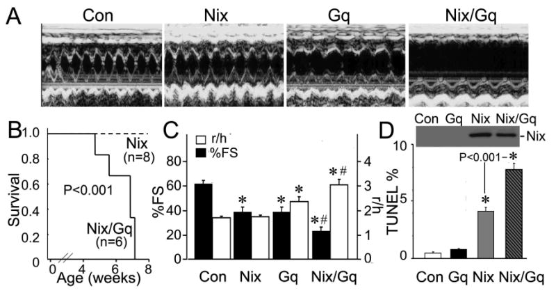
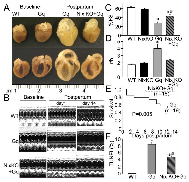
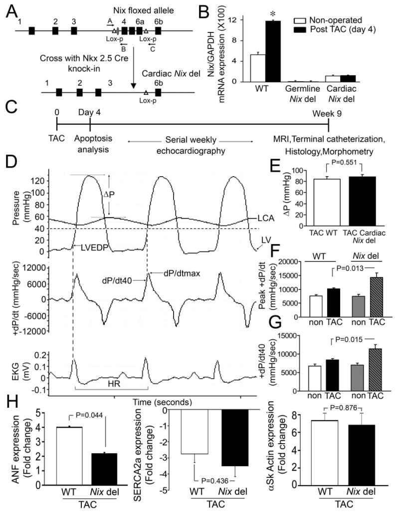
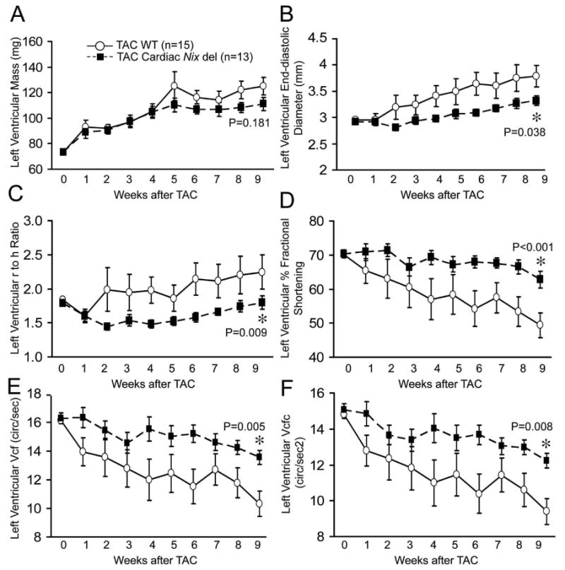
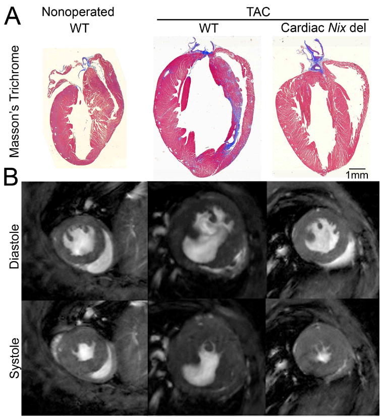
Methods and results: We targeted the gene for proapoptotic Nix, which is transcriptionally upregulated in pressure overload and Gq-dependent hypertrophies, in the mouse germ line or specifically in cardiomyocytes (knockout [KO]) and conditionally overexpressed it in the heart (transgenic [TG]). Conditional forced Nix expression acted synergistically with the prohypertrophic Gq transgene to increase cardiomyocyte apoptosis (0.8+/-0.1% in GqTG versus 7.8+/-0.6% in GqTG+NixTG; P<0.001), causing lethal cardiomyopathy with LV dilation and depressed systolic function (percent fractional shortening, 39+/-4 versus 23+/-4; P=0.042). In the reciprocal experiment, germ-line Nix ablation significantly reduced cardiomyocyte apoptosis (4.8+/-0.2% in GqTG+NixKO versus 8.4+/-0.5% in GqTG; P=0.001), which improved percent fractional shortening (43+/-3% versus 27+/-3%; P=0.017), attenuated LV remodeling, and largely prevented lethality in the Gq peripartum model of apoptotic cardiomyopathy. Cardiac-specific (Nkx2.5-Cre) Nix KO mice subjected to transverse aortic constriction developed significantly less LV dilation by echocardiography and magnetic resonance imaging, maintained concentric remodeling, and exhibited preserved LV ejection fraction (61+/-2% in transverse aortic constriction cardiac Nix KO versus 36+/-6% in transverse aortic constriction wild-type mice; P=0.003) at 9 weeks, with reduced cardiomyocyte apoptosis at day 4 (1.70+/-0.21% versus 2.73+/-0.35%; P=0.032).
Conclusions: Nix-induced cardiomyocyte apoptosis is a major determinant of adverse remodeling in pathological hypertrophies, a finding that suggests therapeutic value for apoptosis inhibition to prevent cardiomyopathic decompensation.
Conflict of interest statement
Conflict of interest disclosures: None
Figures

Comment in
-
Nix: the cardiac Styx between life and death.Circulation. 2008 Jan 22;117(3):338-40. doi: 10.1161/CIRCULATIONAHA.107.750125. Circulation. 2008. PMID: 18212298 Review. No abstract available.
Similar articles
-
USP20 deletion promotes eccentric cardiac remodeling in response to pressure overload and increases mortality.Am J Physiol Heart Circ Physiol. 2024 Nov 1;327(5):H1257-H1271. doi: 10.1152/ajpheart.00329.2024. Epub 2024 Oct 4. Am J Physiol Heart Circ Physiol. 2024. PMID: 39365672
-
Mitochondrial death protein Nix is induced in cardiac hypertrophy and triggers apoptotic cardiomyopathy.Nat Med. 2002 Jul;8(7):725-30. doi: 10.1038/nm719. Epub 2002 Jun 10. Nat Med. 2002. PMID: 12053174
-
Matairesinol blunts adverse cardiac remodeling and heart failure induced by pressure overload by regulating Prdx1 and PI3K/AKT/FOXO1 signaling.Phytomedicine. 2024 Dec;135:156054. doi: 10.1016/j.phymed.2024.156054. Epub 2024 Sep 15. Phytomedicine. 2024. PMID: 39306883
-
Cardiac reanimation: targeting cardiomyocyte death by BNIP3 and NIX/BNIP3L.Oncogene. 2008 Dec;27 Suppl 1:S158-67. doi: 10.1038/onc.2009.53. Oncogene. 2008. PMID: 19641501 Review.
-
Mitochondrial pruning by Nix and BNip3: an essential function for cardiac-expressed death factors.J Cardiovasc Transl Res. 2010 Aug;3(4):374-83. doi: 10.1007/s12265-010-9174-x. Epub 2010 Mar 16. J Cardiovasc Transl Res. 2010. PMID: 20559783 Free PMC article. Review.
Cited by
-
Mitochondria and mitophagy: the yin and yang of cell death control.Circ Res. 2012 Oct 12;111(9):1208-21. doi: 10.1161/CIRCRESAHA.112.265819. Circ Res. 2012. PMID: 23065344 Free PMC article. Review.
-
Involvement and possible role of transglutaminases 1 and 2 in mediating fibrotic signalling, collagen cross-linking and cell proliferation in neonatal rat ventricular fibroblasts.PLoS One. 2023 Feb 27;18(2):e0281320. doi: 10.1371/journal.pone.0281320. eCollection 2023. PLoS One. 2023. PMID: 36848364 Free PMC article.
-
Mitochondrial dynamics and cell death in heart failure.Heart Fail Rev. 2016 Mar;21(2):123-36. doi: 10.1007/s10741-016-9530-2. Heart Fail Rev. 2016. PMID: 26872674 Review.
-
SOCS3 Negatively Regulates Cardiac Hypertrophy via Targeting GRP78-Mediated ER Stress During Pressure Overload.Front Cell Dev Biol. 2021 Jan 26;9:629932. doi: 10.3389/fcell.2021.629932. eCollection 2021. Front Cell Dev Biol. 2021. PMID: 33585485 Free PMC article.
-
Cardioprotective Effects of Adiponectin-Stimulated Autophagy.J Lipid Atheroscler. 2025 Jan;14(1):40-53. doi: 10.12997/jla.2025.14.1.40. Epub 2024 Nov 7. J Lipid Atheroscler. 2025. PMID: 39911962 Free PMC article. Review.
References
-
- Levy D, Garrison RJ, Savage DD, Kannel WB, Castelli WP. Prognostic implications of echocardiographically determined left ventricular mass in the Framingham Heart Study. N Engl J Med. 1990;322:1561–1566. - PubMed
-
- Dorn GW. The fuzzy logic of physiological cardiac hypertrophy. Hypertension. 2007;49:962–970. - PubMed
-
- Massie BM, Schaefer S, Garcia J, McKirnan MD, Schwartz GG, Wisneski JA, Weiner MW, White FC. Myocardial high-energy phosphate and substrate metabolism in swine with moderate left ventricular hypertrophy. Circulation. 1995;91:1814–1823. - PubMed
-
- Diwan A, Dorn GW. Decompensation of cardiac hypertrophy: cellular mechanisms and novel therapeutic targets. Physiology (Bethesda) 2007;22:56–64. - PubMed
Publication types
MeSH terms
Substances
Grants and funding
LinkOut - more resources
Full Text Sources
Other Literature Sources
Molecular Biology Databases
Research Materials
Miscellaneous
