. 2008 Jan;19(1):39-46.
doi: 10.1681/ASN.2006090998.

Inhibition of hypoxia inducible factor hydroxylases protects against renal ischemia-reperfusion injury

Affiliations

Inhibition of hypoxia inducible factor hydroxylases protects against renal ischemia-reperfusion injury

Peter Hill et al. J Am Soc Nephrol. 2008 Jan.

Abstract

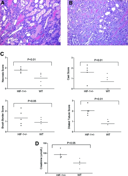
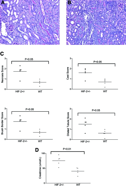
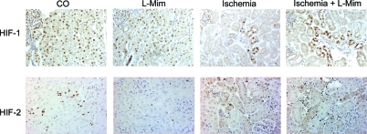
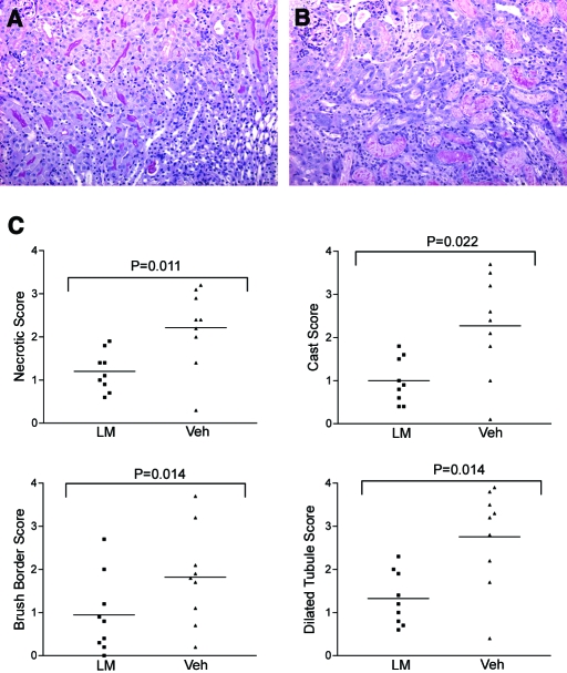
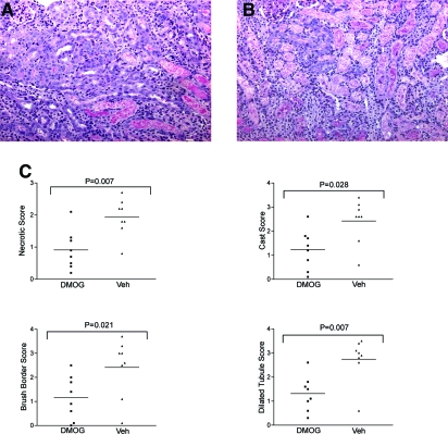
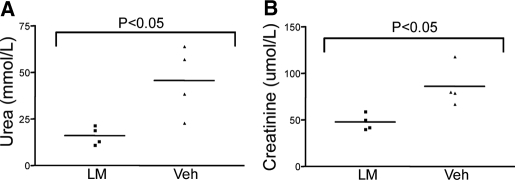
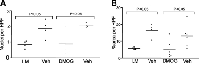
Acute renal failure resulting from hypoperfusion and hypoxia is a significant clinical problem. Hypoxia activates the heterodimeric transcription factor hypoxia inducible factor (HIF), leading to changes in gene expression that promote tissue adaptation and survival. To determine whether HIF may protect the kidney from ischemia-reperfusion injury, we subjected hif1a(+/-) and hif2a(+/-) mice to renal ischemia-reperfusion injury. Injury was substantially more severe in hif(+/-) than in littermate controls, consistent with a protective role for HIF. Because wild-type mice exhibited submaximal HIF accumulation in response to no-flow ischemia, we tested compounds that might augment the protective HIF response following ischemia-reperfusion in these animals. We found that l-mimosine and dimethyloxalylglycine, two small molecules that activate HIF by inhibiting HIF hydroxylases, protected mouse kidneys from ischemia-reperfusion injury. Therefore, pharmacological activation of HIF may offer an effective strategy to protect the kidney from ischemic injury.


Figures

Figure 1.
Figure 1.
Hif1a+/− mice show more severe histologic damage after renal IRI. (A) Representative PAS-stained section after renal ischemia reperfusion from a hif1a+/− mouse. (B) Littermate control. (C) Quantitative scores of tubular injury. (D) Serum creatinine values were significantly higher in hif1a+/− mice compared with littermate controls after bilateral injury. Magnification, ×100.
Figure 2.
Figure 2.
Hif2a+/− mice show more severe histologic damage after renal IRI. (A) Representative PAS-stained section after renal ischemia reperfusion from a hif2a+/− mouse. (B) Littermate control. (C) Quantitative scores of tubular injury. (D) Serum creatinine values were significantly higher in hif2a+/− mice compared with littermate controls after bilateral injury. Magnification, ×100.
Figure 3.
Figure 3.
(Top) HIF-1α immunohistochemistry. (A) After exposure to 0.1% CO, nuclear HIF-1α is detected in epithelial cells throughout the renal cortex. (B) l-Mimosine–treated animals showed widespread activation of HIF-1α. (C) In ischemic mouse kidney, substantially less HIF-1α is detected. (D) Treatment with l-mimosine before ischemic injury increased expression of HIF-1α. In untreated animals, no nuclear signal for HIF-1α was detected (data not shown). (Bottom) HIF-2α immunohistochemistry. (A) 0.1% CO exposure results in HIF-2α in interstitial cells. (B) l-Mimosine treatment leads to HIF-2α activation but less than CO exposure. (C) In ischemic mouse kidney, substantially less HIF-2α is detected. (D) Treatment with l-mimosine before ischemic injury increased expression of HIF-2α. Magnification, ×200.
Figure 4.
Figure 4.
l-Mimosine protects from renal IRI. PAS-stained section from an l-mimosine–treated mouse (A) showing less severe injury compared with section from a vehicle-treated control (B). (C) Tubular injury scores. Magnification, ×100.
Figure 5.
Figure 5.
DMOG protects from renal IRI. (A) PAS-stained section from a DMOG-treated mouse. (B) Vehicle-treated control. (C) Tubular injury scores. Magnification, ×100.
Figure 6.
Figure 6.
Effect of l-mimosine on renal dysfunction after bilateral renal IRI. (A) Serum urea. (B) Serum creatinine. Data are means ± SEM. Significantly lower values were observed in animals that were treated with l-mimosine.
Figure 7.
Figure 7.
Effect of l-mimosine and DMOG on apoptosis and macrophage infiltration after renal IRI. (A) Apoptosis of renal tubular epithelial cells was assessed by TUNEL. Significantly fewer apoptotic nuclei were detected in animals treated with l-mimosine or DMOG. (B) Macrophage infiltration as assessed by CD68 immunohistochemistry was significantly reduced.

References

    1. Maxwell PH: Hypoxia-inducible factor as a physiological regulator. Exp Physiol 90: 791–797, 2005 - PubMed
    1. Wang GL, Jiang BH, Rue EA, Semenza, GL: Hypoxia-inducible factor 1 is a basic-helix-loop-helix-PAS heterodimer regulated by cellular O2 tension. Proc Natl Acad Sci U S A 92: 5510–5514, 1995 - PMC - PubMed
    1. Semenza GL: Surviving ischemia: adaptive responses mediated by hypoxia-inducible factor 1. J Clin Invest 106: 809–812, 2000 - PMC - PubMed
    1. Semenza GL, Wang GL: A nuclear factor induced by hypoxia via de novo protein synthesis binds to the human erythropoietin gene enhancer at a site required for transcriptional activation. Mol Cell Biol 12: 5447–5454, 1992 - PMC - PubMed
    1. Semenza, GL: Targeting HIF-1 for cancer therapy. Nat Rev Cancer 3: 721–732, 2003 - PubMed

Publication types

MeSH terms

LinkOut - more resources