. 2008 Jan;19(1):117-24.
doi: 10.1681/ASN.2007010080.

BMP receptor ALK3 controls collecting system development

Affiliations

BMP receptor ALK3 controls collecting system development

Sunny Hartwig et al. J Am Soc Nephrol. 2008 Jan.

Abstract

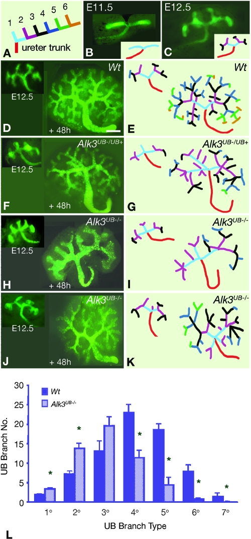
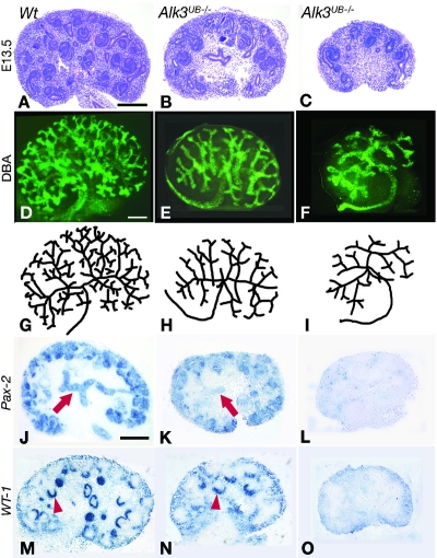
The molecular signals that regulate growth and branching of the ureteric bud during formation of the renal collecting system are largely undefined. Members of the bone morphogenetic protein (BMP) family signal through the type I BMP receptor ALK3 to inhibit ureteric bud and collecting duct cell morphogenesis in vitro. We investigated the function of the BMP signaling pathway in vivo by generating a murine model of ALK3 deficiency restricted to the ureteric bud lineage (Alk3(UB-/-) mice). At the onset of branching morphogenesis, Alk3(UB-/-) kidneys are characterized by an abnormal primary (1 degrees ) ureteric bud branch pattern and an increased number of ureteric bud branches. However, during later stages of renal development, Alk3(UB-/-) kidneys have fewer ureteric bud branches and collecting ducts than wild-type kidneys. Postnatal Alk3(UB-/-) mice exhibit a dysplastic renal phenotype characterized by hypoplasia of the renal medulla, a decreased number of medullary collecting ducts, and abnormal expression of beta-catenin and c-MYC in medullary tubules. In summary, normal kidney development requires ALK3-dependent BMP signaling, which controls ureteric bud branching.


Figures

Figure 1.
Figure 1.
Alk3 is deleted in the ureteric bud lineage of HoxB7Cre;Alk3UB−/− kidneys. (A through C) Expression of Alk3 RNA during early kidney development. (A) Alk3 is expressed along the caudal portion of the Wolffian duct, ventral to the dorsal aorta marked by the arrowhead, and in the nephrogenic mesenchyme (arrow) at E11.5. (B and C) Alk3 is expressed in both the nephrogenic mesenchyme and the ureteric bud at E12.5 and E13.5. Arrows denote the ureteric bud. (D through I) RNA in situ hybridization analysis of Alk3 in E18.5 Wt and Alk3UB−/− kidneys. Boxes in D and E indicate medullary regions shown at higher magnification in F and G; asterisks indicate cortical regions shown at higher magnification in H and I. In Alk3UB−/− kidneys, Alk3 is expressed in nephrogenic mesenchyme but not in collecting ducts (arrows). (J and K) Phospho-Smad1 expression in E11.5 metanephric tissue. Phospho-Smad1 is expressed in a nuclear pattern in ureteric cells in Wt tissue (arrows) (J). Phospho-Smad1 expression is markedly decreased in ureteric cells in Alk3-deficient tissue (arrow) (K). (L) Western blot analysis of E18.5 whole kidney lysates show that ALK3 protein was reduced 57% (P = 0.01), whereas phosphorylated Smad1 (phospho-Smad1) was reduced 53% in Alk3UB−/− kidneys (P = 0.01) (n = 5). Scale bars: (A) 500 μm, (B and C) 25 μm, (D and E) 500 μm, (F through I) 100 μm.
Figure 2.
Figure 2.
Primary ureteric bud branch pattern is disrupted in Alk3UB−/− kidneys. (A) Ureteric bud branches are classified according to their spatial orientation relative to the ureter trunk. (B and C) Green fluorescent protein (GFP) fluorescence in E11.5 and E12.5 Wt kidneys demonstrates the archetypal 1° ureteric bud branch pattern consisting of a ureter trunk (red) connected to two 1° ureteric bud branches (cyan). (D through K) GFP fluorescence of ureteric bud trees at E12.5 and Dolichos biflorus (DBA) fluorescence of these ureteric bud trees after 48 h of culture (+48h), together with corresponding color-coded stick figures. (D and E) The Wt (+48h) ureteric bud tree maintains the 1° branch pattern and exhibits 2° to 6° branch generations. (F and G) Some E12.5 Alk3UB−/UB+ kidneys exhibit a trifid branch pattern (inset in F) that is maintained after 48 h of culture. (H through K) Representative images of the trifid 1° branch pattern observed in the majority of Alk3UB−/− kidneys. (H and I) In some Alk3UB−/− (+48h) kidneys, severe reductions in 3° to 7° branch number and in total ureteric bud branch number are observed. Ureteric bud trunk and tip segments are also dilated, and terminal ureteric bud ampullae formed 4 to 6 new tips. (J and K) Some Alk3UB−/− (+48h) kidneys formed new 1° ureteric bud branches at the junction of the ureter trunk and initial ureteric branches. (L) Quantitation of ureteric bud branch number demonstrates that increased 1° to 2° ureteric bud branch number is associated with decreased 4° to 7° ureteric bud branch number in Alk3UB−/− (+48h) kidneys. Scale bar (D through K) = 100 μm.
Figure 3.
Figure 3.
Histological and molecular phenotype of E13.5 Alk3UB−/− kidneys. (A through C) At E13.5, Alk3UB−/− kidneys exhibited moderate (B) to severe (C) reductions in size and number of mesenchyme- and ureteric bud–derived tissue elements. (D through F) DBA-stained E13.5 Alk3UB−/− kidneys and replica stick figures (G through I) Alk3UB−/− mutants are characterized by a variable decrease in ureteric bud branch number. Some mutants show a moderate decrease (E and H), whereas others demonstrate a severe decrease as well as dilation of ureteric bud tips (F and I). (J through L) Pax2 expression is reduced in the ureteric bud (arrow) in E13.5 moderately affected Alk3UB−/− mutants, whereas in severe mutants Pax2 is reduced in both the ureteric bud and mesenchyme. (M through O) WT-1 expression is markedly reduced in moderately affected mutants (arrowhead) and is undetectable in severe mutants. Scale bars (A through O) = 100 μm.
Figure 4.
Figure 4.
Collecting duct number is reduced in E18.5 Alk3UB−/− kidneys. (A and B) E18.5 Alk3UB−/− kidneys exhibit a paucity of medullary tissue compared with Wt, which suggests a reduction in tubular elements. (C through F) RNA in situ hybridization shows reduced Pax2 expression in Alk3UB−/− collecting ducts. Pax2 mRNA expression is observed in the outer nephrogenic zone (boxed areas) and in tubules extending from the inner medulla to the cortex in Wt kidneys. (C and D) Pax2 mRNA expression is decreased in Alk3UB−/− kidneys. (E and F) Higher power images of the nephrogenic zone demarcated in C and D demonstrate similar levels of Pax2 expression in Wt and Alk3UB−/− kidneys. (G and H) Calbindin28K antibody staining shows marked reduction in collecting duct number (arrows) in Alk3UB−/− kidneys. (I) Collecting duct number is reduced 39% (P = 0.0001) in Alk3UB−/− kidneys. Scale bars: (A through D) 500 μm, (G and H) 100 μm.
Figure 5.
Figure 5.
Adult Alk3UB−/− kidneys exhibit medullary hypoplasia. (A and B) P21 Alk3UB−/− kidneys exhibit cortical cysts, loss of the renal papilla, and medullary hypoplasia. (C and D) Malformation of medullary collecting ducts, identified with anti-calbindin28K antibody, in Alk3UB−/− kidneys (boxed areas in A and B). (E and F) Nuclear phospho-Smad1 expression is not detected in the medulla of Alk3UB−/− kidneys. (G and H) Cytosolic β-catenin expression is increased in Alk3UB−/− kidneys compared with Wt. (I and J) De novo nuclear MYC expression is observed in Alk3UB−/− kidneys. (K) Reduced ALK3 and phospho-Smad1 protein, together with increased β-catenin and MYC proteins, are observed in P21 Alk3UB−/− whole-kidney lysates compared with Wt. Scale bars: (A and B) 1 mm, (C through F) 50 μm, (G and H) 500 μm, (I and J) 50 μm.

References

    1. Saxen L: Organogenesis of the Kidney. Cambridge, Cambridge University Press, 1987
    1. Hogan BL: Bone morphogenetic proteins: Multifunctional regulators of vertebrate development. Genes Dev 10: 1580–1594, 1996 - PubMed
    1. Dudley AT, Robertson EJ: Overlapping expression domains of bone morphogenetic protein family members potentially account for limited tissue defects in BMP7 deficient embryos. Dev Dyn 208: 349–362, 1997 - PubMed
    1. Dewulf N, Verschueren K, Lonnoy O, Morén A, Grimsby S, Vande Spiegle K, Miyazono K, Huylebroeck D, Ten Dijke P: Distinct spatial and temporal expression patterns of two type 1 receptors for bone morphogenetic proteins during mouse embryogenesis. Endocrinology 136: 2652–2663, 1995 - PubMed
    1. Piscione TD, Yager TD, Gupta IR, Grinfeld B, Pei Y, Attisano L, Wrana JL, Rosenblum ND: BMP-2 and OP-1 exert direct and opposite effects on renal branching morphogenesis. Am J Physiol 273: F961–F975, 1997 - PubMed

Publication types

Substances

LinkOut - more resources