. 2008 Jan;19(1):125-34.
doi: 10.1681/ASN.2007030261.

Hypokalemic nephropathy is associated with impaired angiogenesis

Affiliations

Hypokalemic nephropathy is associated with impaired angiogenesis

Sirirat Reungjui et al. J Am Soc Nephrol. 2008 Jan.

Abstract

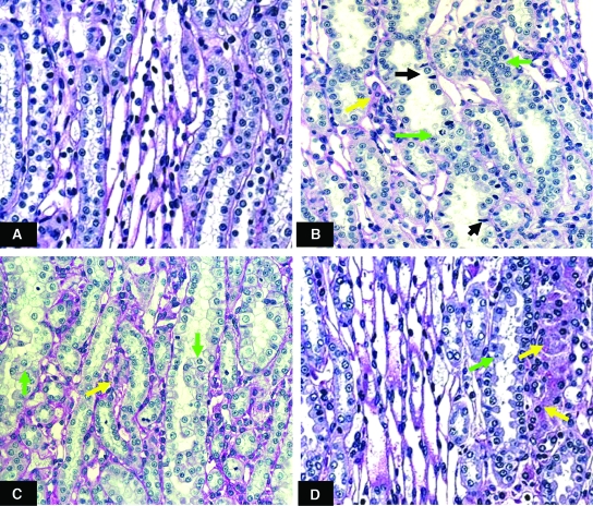
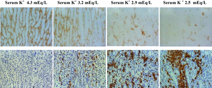
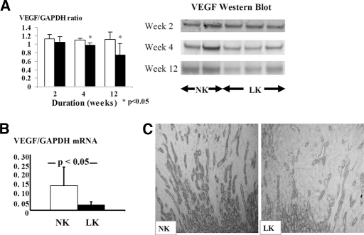
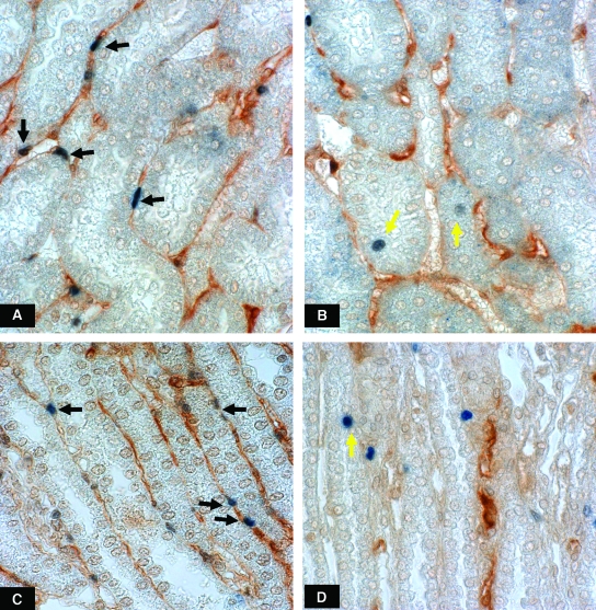
Hypokalemic nephropathy is associated with alterations in intrarenal vasoactive substances, leading to vasoconstriction, salt-sensitivity, and progression of interstitial fibrosis. In this study, we investigated whether hypokalemic nephropathy might also involve impaired renal angiogenesis. Sprague-Dawley rats that were fed low-potassium diets developed peritubular capillary loss that began in the inner stripe of the outer medulla (week 2) and progressed to the outer stripe of the outer medulla (week 4) and cortex (week 12). These changes were associated with increased macrophage infiltration, increased expression of both monocyte chemoattractant protein-1 and TNF-alpha, and a loss of vascular endothelial growth factor and endothelial nitric oxide synthase. Renal thiobarbituric acid-reactive substances, markers of oxidative stress, were increased late in disease. In conclusion, hypokalemic nephropathy is associated with impaired renal angiogenesis, evidenced by progressive capillary loss, reduced endothelial cell proliferation, and loss of VEGF expression.


Figures

Figure 1.
Figure 1.
Periodic acid-Schiff reagent staining within ISOM of normal K+ rats (A) and low K+ rats at weeks 2 (B), 4 (C) and 12 (D). Magnification 400x. Hypokalemic rats developed renal tubular cell hypertrophy and hyperplasia (green arrows) and interstitial cell infiltration (yellow arrows). Pyknotic nuclei representing apoptotic cells were demonstrated in renal tubular cells at week 2 (black arrows).
Figure 2.
Figure 2.
Proliferating cell nuclear antigen (PCNA; upper panel) and osteopontin (lower panel) immunostaining within the ISOM area of normal K+ (NK) and low K+ rats (LK) at weeks 2, 4 and 12. Magnification 400x. Hypokalemic rats had more renal tubular cell proliferation as shown with PCNA positive cells especially at week 2. Renal tubular damage represented by osteopontin staining was progressive in hypokalemic rats.
Figure 3.
Figure 3.
Renal MCP-1 concentration and ED-1 immunostaining within ISOM area of normal K+ (NK, represented with white bars) and low K+ rats (LK, represented with black bars) at weeks 2, 4 and 12. Magnification 630x. Renal MCP-1 content progressively increased in hypokalemic rats which was significantly higher at week 12 compared with week 2 (P = 0.01) and positively correlated with the density of macrophage infiltration which also increased with time.
Figure 4.
Figure 4.
JG-12 immunostaining within the ISOM area of normal K+ (A) and low K+ rats at weeks 2 (B), 4 (C) and 12 (D). Magnification 400x. The reduction in peritubular capillaries was progressive over time.
Figure 5.
Figure 5.
Correlation between peritubular capillary loss and macrophage infiltration within the ISOM area at week 12. Peritubular capillaries are represented by JG-12 staining (upper panel) and macrophage infiltration by ED-1 staining (lower panel). The severity of both peritubular capillary loss and macrophage infiltration correlated with each other as well as with the level of serum K+. Magnification 400x.
Figure 6.
Figure 6.
Reduction of renal VEGF mRNA and protein expressions in the hypokalemic rats. (A) VEGF protein represented by Western Blot which was reduced at weeks 4 and 12 of hypokalemic nephropathy (normal K+ rats; NK represented with white bars and low K+ rats; LK represented with black bars). (B) VEGF mRNA expression at week 12 of low K+ diet (C) Renal tubular VEGF expression at week 12 of the study demonstrated by immunohistochemistry (200x)
Figure 7.
Figure 7.
Endothelial cell proliferation, represented by positive double staining of endothelial cells with thrombomodulin and PCNA (black arrows) within OSOM area of the normal K+ rat (A) and low K+ rat at week 4 (B) and within the ISOM area of normal K+ rat (C) and low K+ rat at week 12 (D). Hypokalemic rats had reduced numbers of proliferating endothelial cells. In contrast, proliferating renal tubular cells were increased in hypokalemic rats (yellow arrows). Magnification 630x.
Figure 8.
Figure 8.
Rats fed low K diet have decreased renal eNOS expression at weeks 4 and 12. (A) Renal eNOS mRNA expression at week 12 of the study. (B) eNOS protein represented by Western Blot was reduced in hypokalemic rats at weeks 4 and 12 (C) Immunohistochemical staining of eNOS antibody showed reduced eNOS expression demonstrated in peritubular capillaries within ISOM area at week 12 (Magnification 400x).

Similar articles

Cited by

References

    1. Tolins JP, Hostetter MK, Hostetter TH: Hypokalemic nephropathy in the rat: Role of ammonia in chronic tubular injury. J Clin Invest 79: 447–458, 1987 - PMC - PubMed
    1. Amlal H, Krane CM, Chen Q, Soleimani M: Early polyuria and urinary concentrating defect in potassium deprivation. Am J Physiol Renal Physiol 279: F655–F663, 2000 - PubMed
    1. Fourman P, McCance RA, Parker RA: Chronic disease in rats following a temporary deficiency of potassium. Br J Exp Pathol 37: 40–44, 1956 - PMC - PubMed
    1. Kimura T, Nishino T, Maruyama N, Hamano K, Kubo A, Iwano M, Shiiki H: Expression of Bcl-2 and Bax in hypokalemic nephropathy in rats. Pathobiology 69: 237–248, 2001 - PubMed
    1. Tsao T, Fawcett J, Fervenza FC, Hsu FW, Huie P, Sibley RK, Rabkin R: Expression of insulin-like growth factor-1 and transforming growth factor-beta in hypokalemic nephropathy in the rat. Kidney Int 59: 96–105, 2001 - PubMed

Publication types