. 2008 Jan 15;180(2):1098-106.
doi: 10.4049/jimmunol.180.2.1098.

CXCL9 and CXCL10 expression are critical for control of genital herpes simplex virus type 2 infection through mobilization of HSV-specific CTL and NK cells to the nervous system

Affiliations

CXCL9 and CXCL10 expression are critical for control of genital herpes simplex virus type 2 infection through mobilization of HSV-specific CTL and NK cells to the nervous system

Manoj Thapa et al. J Immunol. .

Abstract

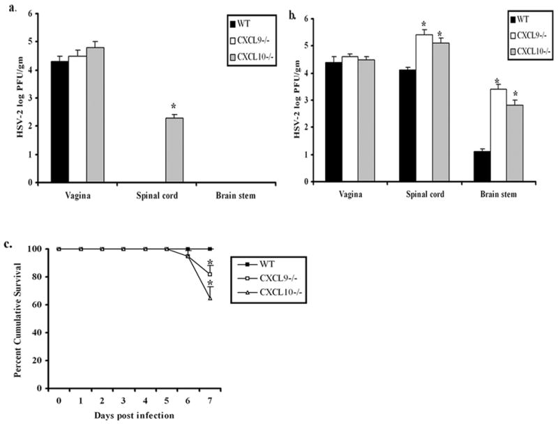
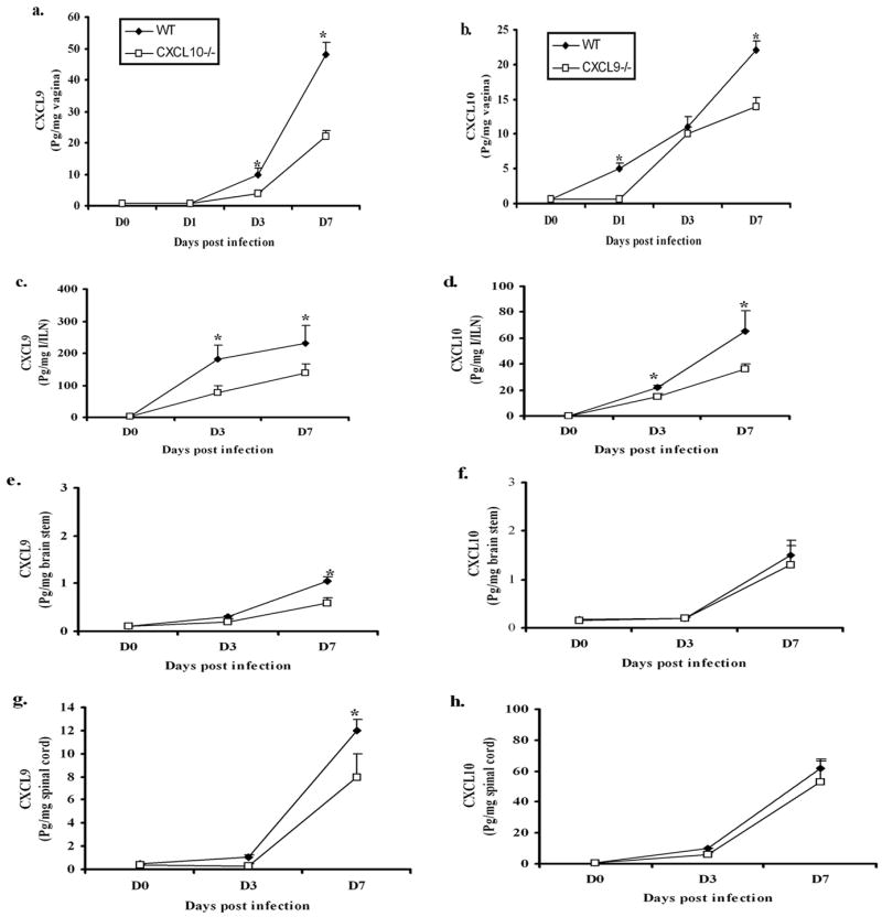
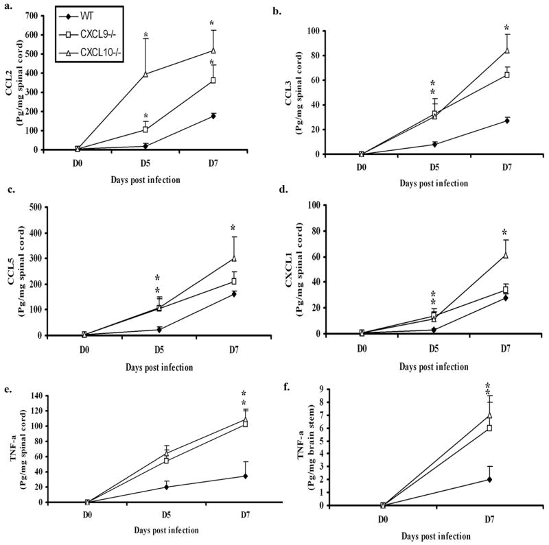
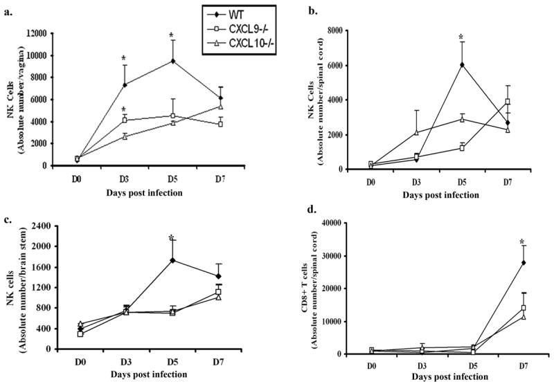
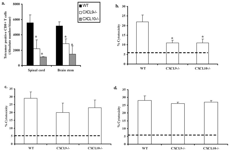
CXCL9 and CXCL10 mediate the recruitment of T lymphocytes and NK cells known to be important in viral surveillance. The relevance of CXCL10 in comparison to CXCL9 in response to genital HSV-2 infection was determined using mice deficient in CXCL9 (CXCL9-/-) and deficient in CXCL10 (CXCL10-/-) along with wild-type (WT) C57BL/6 mice. An increased sensitivity to infection was found in CXCL10-/- mice in comparison to CXCL9-/- or WT mice as determined by detection of HSV-2 in the CNS at day 3 postinfection. However, by day 7 postinfection both CXCL9-/- and CXCL10-/- mice possessed significantly higher viral titers in the CNS in comparison to WT mice consistent with mortality (18-35%) of these mice within the first 7 days after infection. Even though CXCL9-/- and CXCL10-/- mice expressed elevated levels of CCL2, CCL3, CCL5, and CXCL1 in the spinal cord in comparison to WT mice, there was a reduction in NK cell and virus-specific CD8+ T cell mobilization to this tissue, suggesting CXCL9 and CXCL10 are critical for recruitment of these effector cells to the spinal cord following genital HSV-2 infection. Moreover, leukocytes from the spinal cord but not from draining lymph nodes or spleens of infected CXCL9-/- or CXCL10-/- mice displayed reduced CTL activity in comparison to effector cells from WT mice. Thus, the absence of CXCL9 or CXCL10 expression significantly alters the ability of the host to control genital HSV-2 infection through the mobilization of effector cells to sites of infection.


Figures

Figure 1
Figure 1
CXCL9−/− and CXCL10−/− mice are highly susceptible to HSV-2 genital infection. Wild type (WT), CXCL9−/− and CXCL10−/− mice (n=6 mice/group) were infected with HSV-2 (2000 pfu/vagina). At indicated times day 3 pi (a), and day 7 pi (b), mice were exsanguinated and vaginal tissue, brain stem and spinal cord were processed and assayed for viral titer by standard plaque assay and viral titer is expressed as mean log PFU ± SEM. (c), WT, CXCL9−/− and CXCL10−/− mice (n=44–59 mice/group) were infected with HSV-2 (2000 pfu/vagina) and were monitored and recorded for survival. The bars represent the mean ± SEM summarizing the results of three independent experiments. *, p<.05 comparing WT to CXCL9−/− and CXCL10−/− mice.
Figure 2
Figure 2
CXCL9 & CXCL10 levels are elevated in tissue specific manner. WT, CXCL9−/− and CXCL10−/− (n=6/group) were infected with HSV-2 (2000 pfu/vagina). At indicated times, the mice were exsanguinated and vaginal tissue (a & b), Inguinal/Iliac lymph node (I/ILN) (c & d), brain stem (e & f), and spinal cord (g & h) were removed, processed and assayed for CXCL9 (left panel) and CXCL10 (right panel) content using ELISA. The weight of the tissue was used to normalize the amount of chemokine per milligram of tissue (expressed as pg per mg of tissue). Day 0 time point represents uninfected controls. Each point represents the mean ± SEM summarizing the results of two independent experiments. *, p<.05 comparing WT to CXCL9−/− and CXCL10−/− groups.
Figure 3
Figure 3
Chemokine/cytokine levels are elevated in the vaginal tissue of CXCL10−/−mice. WT, CXCL9−/−, CXCL10−/− mice (n=6/group) were infected with HSV-2 (2000 pfu/vagina). At indicated times, the mice were exsanguinated and vaginal tissues were removed, processed and assessed for CCL2, CCL3, CCL5, CXCL1, TNF-α, and IFN-and IL-12p70 (not shown) content using a suspension array system and ELISA. Samples were analyzed in duplicate along with standard provided to generate standard curves for each analyte. The weight of the tissue was used to normalize amount of cytokine/chemokine per milligram of tissue weight. Day 0 time point represents uninfected controls. Each point represents the mean ± SEM summarizing the results of two independent experiments. *, p<.05 comparing the WT to CXCL9−/− and CXCL10−/−groups.
Figure 4
Figure 4
Chemokine/cytokine levels are elevated in the nervous system of chemokine knockout mice. WT, CXCL9−/− , CXCL10−/− mice (n=6/group) were infected with HSV-2 (2000 pfu/vagina). At indicated times, the mice were exsanguinated and the spinal cords and brain stems were removed, processed and assessed for CCL2, CCL3, CCL5, CXCL1 and TNF-α content using a suspension array system and ELISA. As there were no differences found in other analytes measured, only TNF-α level is shown for brain stem. Samples were analyzed in duplicate along with standard provided to generate standard curves for each analyte. The weight of the tissue was used to normalize amount of cytokine/chemokine per milligram of tissue weight. Day 0 time point represents uninfected controls. Each point represents the mean ± SEM summarizing the results of two independent experiments. *, p<.05 comparing the WT to CXCL9−/− and CXCL10−/−groups.
Figure 5
Figure 5
NK and CD8+ T cells infiltration into infected tissue of chemokine knockout mice is reduced or delayed. WT, CXCL9−/− and CXCL10−/− mice (n= 6/group) were infected with HSV-2 (2000 pfu/vagina) and subsequently exsanguinated at indicated times post infection (pi) and (a) vaginal tissue, (b) spinal cord, and (c) brain stem samples were processed and analyzed for NK cell (NK1.1+CD3CD45high) content by flow cytometry. (d) Similarly, spinal cords were processed and analyzed for CD8+ T (CD3+CD8+CD45high) cells content using flow cytometry. Day 0 time point represents uninfected controls. Each point represents the mean ± SEM summarizing the results of three independent experiments. *, p<0.05 comparing WT to CXCL9−/− and CXCL10−/−.
Figure 6
Figure 6
Reduced HSV-2 specific CD8+ T cell recruitment is associated with decreased cytolytic activity. WT, CXCL9−/− and CXCL10−/− mice were (n= 6/group) were infected with HSV-2 (2000 pfu/vagina). At indicated times, mice were exsanguinated and spinal cord, Spleen and I/ILN were processed and analyzed (a), for tetramer positive T cells at day 7 and (b), for CTL activity using percoll gradient enriched SC leukocytes, (c), for CTL activity using I/ILN lymph node cells and (d), for CTL activity using spleen cells. For tetramer staining, MHC I tetramer specific for HSV peptide gB498–505 (SSIEFARL) was used to assay the total tetramer positive cells in the spinal cord. For CTL assay, HSV-2 infected target cells (MC57G) were labeled with CFSE dye and incubated with percoll gradient enriched SC leukocytes or spleen cells or lymph node cells at a E:T ratio of 10:1 for 4 hr at 37°C. Propidium iodide was added after the 4 hr incubation and percent lysis was determined using flow cytometry. The line in b-d indicates the background PI incorporation into CSFE-labeled targets incubated only or in the presence of spleen cells from uninfected mice. The bars represent the mean ± SEM summarizing the results of two independent experiments. *, p<.05 comparing WT to CXCL9−/− and CXCL10−/−groups.

Similar articles

Cited by

References

    1. Inagaki K, Daikoku T, Goshima F, Nashiyama Y. Impaired induction of protective immunity by highly virulent herpes simplex type 2 in a murine model of genital herpes. Arch Virol. 2000;145:1989–2002. - PubMed
    1. Harandi AM, Svennerholm B, Holmgren J, Eriksson K. Differential roles of B cells and IFN- -secreting CD4+ T cells in innate and adaptive immune control of genital herpes simplex virus type 2 infection in mice. J Gen Virol. 2001;82:845–853. - PubMed
    1. Whitley RJ, Miller RL. Immunologic approach to Herpes Simplex Virus. Viral Immunol. 2001;14(2):111–118. - PubMed
    1. Duerst RJ, Morrison LA. Innate immunity to herpes simplex virus type 2. Viral Immunol. 2003;16(4):475–490. - PubMed
    1. MasCasullo V, Fam E, Keller MJ, Herold BC. Role of mucosal immunity preventing genital herpes infection. Viral Immunol. 2005;18(4):595–606. - PubMed

Publication types