Interleukin-6 concentrations in wound fluids rather than serological markers are useful in assessing bacterial triggers of ulcer inflammation
- PMID: 18179556
- PMCID: PMC7951795
- DOI: 10.1111/j.1742-481X.2007.00347.x
Interleukin-6 concentrations in wound fluids rather than serological markers are useful in assessing bacterial triggers of ulcer inflammation
Abstract
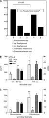
Bacterial pathogenicity, microbial load and diversity are decisive for outcome and therapy of non healing ulcers. However, until now, no routine laboratory parameter is available to assess the inflammatory level caused by chronic wound infections. We thus investigated the usefulness of levels of interleukin (IL)-6 and tumour necrosis factor alpha (TNFalpha) in wound fluids for assessing ulcer inflammation in the presence or absence of microbial triggers. In addition, the predictive values of local cytokine analyses were compared with those of C-reactive protein (CRP) and lipopolysaccharide-binding protein (LBP) because serological markers are normally used to underline the suspicion of wound infections. The present data from chronic arterial and venous ulcers (n = 45) clearly show that mixed bacterial infections increased IL-6 and TNFalpha concentrations in wound fluids when compared with ulcers with a monomicrobial infection (P < 0.01 and P < 0.05, respectively). IL-6 was also significantly elevated when a high bacterial load [versus <10(5) colony-forming units (cfu)/ml: P = 0.04] or an infection with Pseudomonas was observed (versus isolation of non Pseudomonas strains: P = 0.05). Although distinct proinflammatory triggers may interfere with regard to cytokine levels, sensitivity and specificity were significant in predicting bacterial risk factors, particularly for IL-6 at a designated cut-off of 125 pg/ml (sensitivity and specificity for predicting a mixed infection: 70% and 64%, for predicting a bacterial load of >10(5) cfu/ml: 62% and 57% and for predicting an infection with Pseudomonas: 90% and 57%). In contrast to local cytokine levels, the serological markers CRP and LBP were not associated with the presence of any of the investigated bacterial triggers. Focusing on the aim of the study, IL-6 analysed in wound washouts seems to be a useful diagnostic marker for a sensitive and specific assessment of ulcers inflammation with regard to bacterial triggers.
Figures

References
-
- Vermeulen H, Schreuder S, Lubbers M, Ubbink D. Inter‐ and intraobserver agreement among doctors and nurses in the classification of open wounds. J Wound Healing 2005:V40–2.(abstract), ETRS European Wound Conference.
-
- Rotstein OD, Pruett TL, Simmons RL. Mechanisms of microbial synergy in polymicrobial surgical infections. Rev Infect Dis 1985;7:151–70. - PubMed
-
- Serralta VW, Harrison‐Balestra C. Lifestyles of bacteria in wounds: presence of biofilms? Wounds Compend Clin Res Pract 2001;13:29–34.
-
- Robson MC. Wound infection: a failure of wound healing caused by an imbalance of bacteria. Surg Clin North Am 1997;77:637–50. - PubMed
-
- Heggers JP. Defining infection in chronic wounds: does it matter? Wound Care 1998;7:389–92. - PubMed
Publication types
MeSH terms
Substances
LinkOut - more resources
Full Text Sources
Other Literature Sources
Medical
Research Materials
Miscellaneous

