. 2008 Mar;36(3):350-9.
doi: 10.1016/j.exphem.2007.10.007. Epub 2008 Jan 7.

Derivation and immunological characterization of mesenchymal stromal cells from human embryonic stem cells

Affiliations

Derivation and immunological characterization of mesenchymal stromal cells from human embryonic stem cells

Parul Trivedi et al. Exp Hematol. 2008 Mar.

Abstract

Objective: We have previously shown the simultaneous generation of CD73(+) mesenchymal stromal cells (MSCs) along with CD34(+) hematopoietic cells from human embryonic stem cells (ESCs) when they are cocultured with OP9 murine stromal cells. We investigated whether MSCs can be derived from human ESCs without coculturing with OP9 cells, and if such cells exhibit immunological properties similar to MSCs derived from adult human bone marrow (BM).

Materials and methods: Our starting populations were undifferentiated human ESCs cultured on Matrigel-coated plates without feeder cells. The differentiated fibroblast-looking cells were tested for expression of MSC markers and their potential for multilineage differentiation. We investigated surface expression of human leukocyte antigen (HLA) molecules on these MSCs before and after treatment with interferon-gamma (IFN-gamma). We also tested the proliferative response of T-lymphocytes toward MSCs and the effects of MSCs in mixed lymphocyte reaction (MLR) assays.

Results: We derived populations of MSCs from human ESCs with morphology, cell surface marker characteristics, and differentiation potential similar to adult BM-derived MSCs. Similar to BM-derived MSCs, human ESC-derived MSCs express cell surface HLA class I (HLA-ABC) but not HLA class II (HLA-DR) molecules. However, stimulation with IFN-gamma induced the expression of HLD-DR molecules. Human ESC-derived MSCs did not induce proliferation of T-lymphocytes when cocultured with peripheral blood mononuclear cells. Furthermore, ESC-derived MSCs suppressed proliferation of responder T-lymphocytes in MLR assays.

Conclusions: MSCs can be derived from human ESCs without feeder cells. These human ESC-derived MSCs have cell surface markers, differentiation potentials, and immunological properties in vitro that are similar to adult BM-derived MSCs.


Figures

Figure-1
Figure-1
The majority of undifferentiated human embryonic stem cells (ESCs) (graph-A) are positive for the SSEA-4 marker but they do not express the CD73 marker, one of the markers expressed on mesenchymal stromal cells (MSCs). Progressively during the MSC differentiation process human ESCs loose their SSEA-4 marker and become positive for CD73 (graphs-B and C). Fluorescent activated cell sorting analysis of passage-1 human ESC-derived MSCs shows that they do not express the SSEA-4 marker, but are uniformly positive for the CD73 marker (graph-D).
Figure-2
Figure-2
Upper panels show representative microscopic views of human embryonic stem cells (ESC)-derived mesenchymal stromal cells (MSCs) from H1, H7 and H9 cell lines. The middle panels show a normal karyotype for each human ESC-derived MSC line when tested at passage 4-5 after derivation. The lower panels show the differentiation of H9-ESC-derived MSCs into osteogenic (lower-left), adipogenic (lower-middle), and chondrogenic (lower-right) lineages. Von Kossa staining shows deposits of calcium crystals (lower left), Oil red O staining shows lipid vacuoles (lower middle), and Safranin O staining shows cartilage-specific glycosaminoglycans (lower right), respectively. All photomicrographs of MSCs or their differentiated progenies were taken with a Leica DFC320 digital camera on a Leica DM IRB microscope using C Plan 20×0.22 objective except the chondrogenic picture that was taken by N plan 10×0.12 objective (lower right).
Figure-3
Figure-3
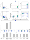
Panel-A shows representative fluorescent activated cell sorting analysis of H1-MSCs, H7-MSCs, H9-MSCs, BM-MSC-1215, and BM-MSC-5066R for different cell surface markers. Table-B shows the mean value (± standard error) of the expression of corresponding markers from at least three sets of experiments.
Figure-4
Figure-4
Human embryonic stem cells (ESC)-derived mesenchymal stromal cells (MSCs) or bone marrow (BM)-derived MSCs express HLA-ABC but do not express HLA-DR. Treatment of human ESC-derived MSCs with IFN-γ induces the cell surface expression of HLA-DR but to a lesser degree compared to that seen from stimulated BM-derived MSCs.
Figure-5
Figure-5
A: Upper panel shows one representative carboxyfluorescein diacetate succinimidyl ester (CFSE) assay showing the T-lymphocyte fraction of peripheral blood mononuclear cells (PB-MNCs) does not proliferate when they are cultured alone. Lower panels show T-lymphocytes labeled with CFSE dividing in response to third-party PB-MNCs. Proliferation index (PI) is calculated using FlowJo software. B: Mean value for proliferation index (± standard error based on at least three sets of experiments) of responder T-lymphocytes cultured alone, co-cultured with non-stimulated or PHA-stimulated PB-MNCs, and human embryonic stem cell-derived or bone marrow-derived mesenchymal stromal cells (MSCs) are shown alongside a representative CFSE assay. MSCs do not induce the proliferation of T-lymphocytes.
Figure-6
Figure-6
Mesenchymal stromal cell (MSCs) cultured in direct contact or in the presence of a semi-permeable membrane in transwells, and also when their conditioned media (CM) were added to mixed lymphocyte reactions (MLRs) suppressed the proliferation of responder T-lymphocytes towards stimulator cells (pooled peripheral blood mononuclear cells). Negative controls included carboxyfluorescein diacetate succinimidyl ester (CFSE) stained T-lymphocytes alone and positive controls included MLRs performed in the absence of MSCs or CM. Culturing in direct contact was always more immunosuppressive compared to transwell culture (this difference did not reach statistical significance for H9-MSCs and BM-MSC-5066R) and compared to CM (this difference did not reach statistical significance for H9-MSCs).

References

    1. Friedenstein AJ, Chailakhyan RK, Latsinik NV, Panasyuk AF, Keiliss-Borok IV. Stromal cells responsible for transferring the microenvironment of the hemopoietic tissues. Cloning in vitro and retransplantation in vivo. Transplantation. 1974;17:331–340. - PubMed
    1. Caplan AI. Mesenchymal stem cells. JOrthopRes. 1991;9:641–650. - PubMed
    1. Pittenger MF, Mackay AM, Beck SC, et al. Multilineage potential of adult human mesenchymal stem cells. Science. 1999;284:143–147. - PubMed
    1. Javazon EH, Beggs KJ, Flake AW. Mesenchymal stem cells: paradoxes of passaging. ExpHematol. 2004;32:414–425. - PubMed
    1. Seeberger KL, Dufour JM, Shapiro AM, Lakey JR, Rajotte RV, Korbutt GS. Expansion of mesenchymal stem cells from human pancreatic ductal epithelium. Laboratory investigation; a journal of technical methods and pathology. 2006;86:141–153. - PubMed

Publication types

MeSH terms