

doi: 10.1016/j.ajhg.2007.08.005.
SERKAL syndrome: an autosomal-recessive disorder caused by a loss-of-function mutation in WNT4
Affiliations
- PMID: 18179883
- PMCID: PMC2253972
- DOI: 10.1016/j.ajhg.2007.08.005
Item in Clipboard
SERKAL syndrome: an autosomal-recessive disorder caused by a loss-of-function mutation in WNT4
Am J Hum Genet.
2008 Jan.
Abstract
The WNT-signaling pathway plays a major role during mammalian embryogenesis. We report a novel autosomal-recessive syndrome that consists of female to male sex reversal and renal, adrenal, and lung dysgenesis and is associated with additional developmental defects. Using a candidate-gene approach, we identified a disease-causing homozygous missense mutation in the human WNT4 gene. The mutation was found to result in markedly reduced WNT4 mRNA levels in vivo and in vitro and to downregulate WNT4-dependent inhibition of beta-catenin degradation. Taken together with previous observations in animal models, the present data attribute a pivotal role to WNT4 signaling during organogenesis in humans.
Figures
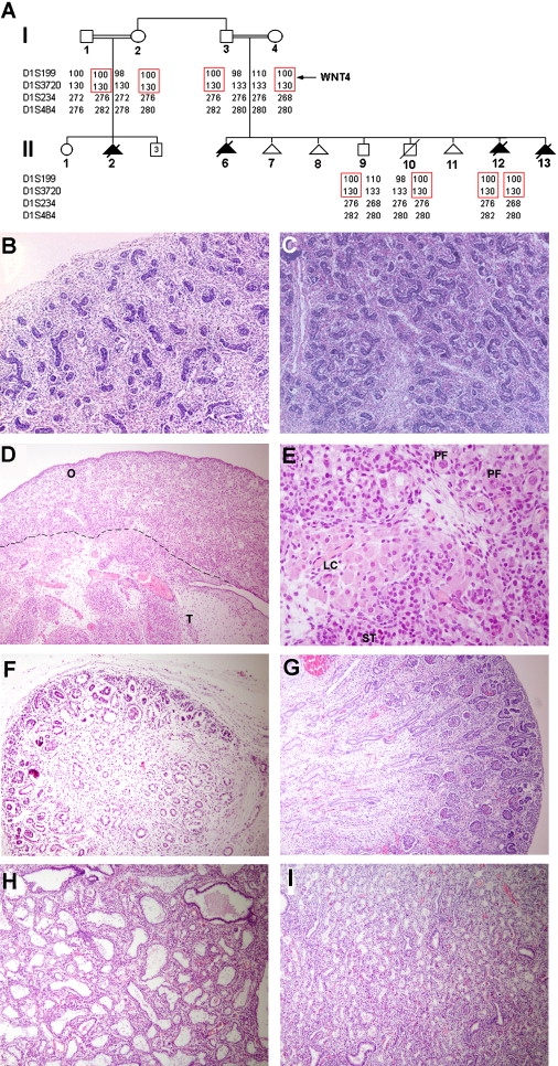
Pedigree Structure and Pathological Features (A) Family pedigree and haplotype analysis using polymorphic microsatellite markers on 1p36.23-p35.1. The disease-associated haplotype is boxed in red. The position of the WNT4 gene is indicated. (B–I) Histopathogical examination of tissues obtained from affected fetuses (B, D, F, and H) and from a normal 19-week-old fetus (C, E, G, and I). Affected 46,XX fetus II-12 displays (B) a normal-looking testis (sex reversal) with coiled seminiferous tubules and numerous Leydig cells throughout the interstitium as compared with the testis of an 46,XY healthy fetus (C); affected fetus II-6 demonstrates both testicular (“T”) and ovarian (“O”) tissues (D) including seminiferous tubules (“ST”), Leydig cells (“LC”), and primordial follicles (“PF”) (E); the kidney of affected fetus II-12 (F) showed a very thin rim of nephrogenic zone, with decreased numbers of mature glomeruli, and very few proximal tubules with immature interstitial tissue resembling undifferentiated mesenchyme of dysplastic kidney as compared with a normal fetal kidney at 19 weeks of gestation (G), where the numerous glomeruli identify the renal cortex; examination of lung tissues of affected fetus II-2 (H) revealed focal marked dilatation of distal airways compared with control fetal lung tissue (I) (magnification, 50× [B and C], 100× [D, F, G, H, and I], 400× [E]).

Mutation Analysis (A) Sequence analysis revealed in an affected fetus (II-12) a homozygous transition (c.C341T) (blackened letter, upper panel), resulting in amino acid substitution A114V. The wild-type sequence is given for comparison (lower panel). (B) c.C314T creates a novel recognition site for HpyCH4IV endonuclease. A 699 bp fragment was amplified as described in Material and Methods and digested with HpyCH4IV. The affected fetus displays two fragments, of 172 bp and 527 bp, that are carried in a heterozygous state by all other family members. (C) The amino acid (upper panel) and cDNA (lower panel) sequences of human WNT4 were compared to those of other human WNT proteins and WNT4 orthologs in other species. Note the high degree of conservation of both amino acid A114 (upper panel) and nucleotide C341 (lower panel).
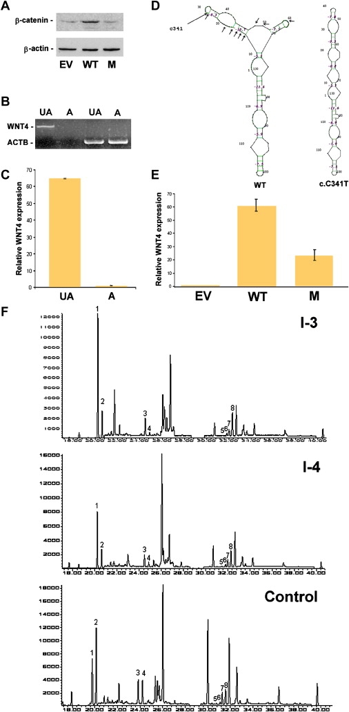
Consequences of Mutation c.C341T (A) Western-blot analysis of β-catenin and β-actin expression in OVCAR3 cells transfected with empty vector (EV), wild-type WNT4 cDNA (WT), and c.C341T mutant cDNA (M). (B) Expression of WNT4 and ACTB, coding for β-actin, assessed with RT-PCR in amniocytes obtained from an affected embryo (“A”) as compared with control amniocytes (“UA”). (C) Quantitative real-time PCR analysis for gene expression of WNT4 in amniocytes obtained from an affected embryo (“A”) as compared with control amniocytes (“UA”) (bars represent standard errors). Expression levels are expressed as absolute values normalized relative to ACTB RNA levels. (D) Predicted secondary structures of wild-type (“WT”) and c.C314T mutant WNT4 RNA spanning exon 3. The position of c.C341T and mutations resulting in a similar change in mRNA secondary structure are marked with arrows. (E) Quantitative real-time PCR analysis for gene expression of WNT4 in OVCAR3 cells transfected with empty vector (“EV”), wild-type WNT4 cDNA (“WT”), and c.C341T mutant cDNA (“M”). Expression levels are expressed as absolute values normalized relative to ACTB RNA levels (bars represent standard errors). (F) Chromatograms (GC-MS, SIM mode) of urinary-steroid hormones. In both parents, 5-α/5-β isomer ratios [Androsterone/Etiocholanolone(1,2);11-OH-Androsterone/11-OH-etiocholanolone (3,4); α-tetrahydrocorticosterone (THB)/β-THB (5,6); α-tetrahydrocortisol (THF)/β-THF (7,8)], reveal an excess of activity of the enzyme 5-alpha-reductase. The 5α-isomers are 1, 3, 6, and 8; the 5β-isomers are 2, 4, 5, and 7.
References
MeSH terms
Substances
LinkOut - more resources
Full Text Sources
Other Literature Sources
Medical
Molecular Biology Databases
