. 2008 Jan;82(1):73-80.
doi: 10.1016/j.ajhg.2007.09.002.

Oncostatin M receptor-beta mutations underlie familial primary localized cutaneous amyloidosis

Affiliations

Oncostatin M receptor-beta mutations underlie familial primary localized cutaneous amyloidosis

Ken Arita et al. Am J Hum Genet. 2008 Jan.

Abstract

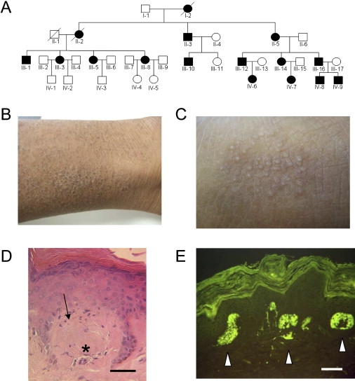
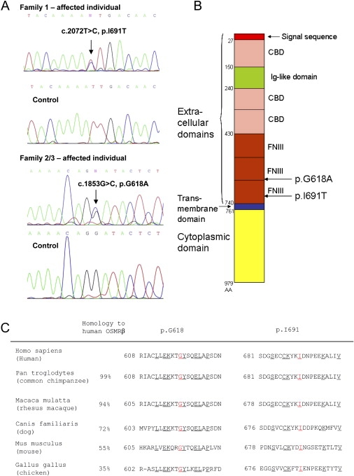
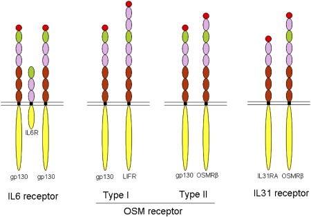
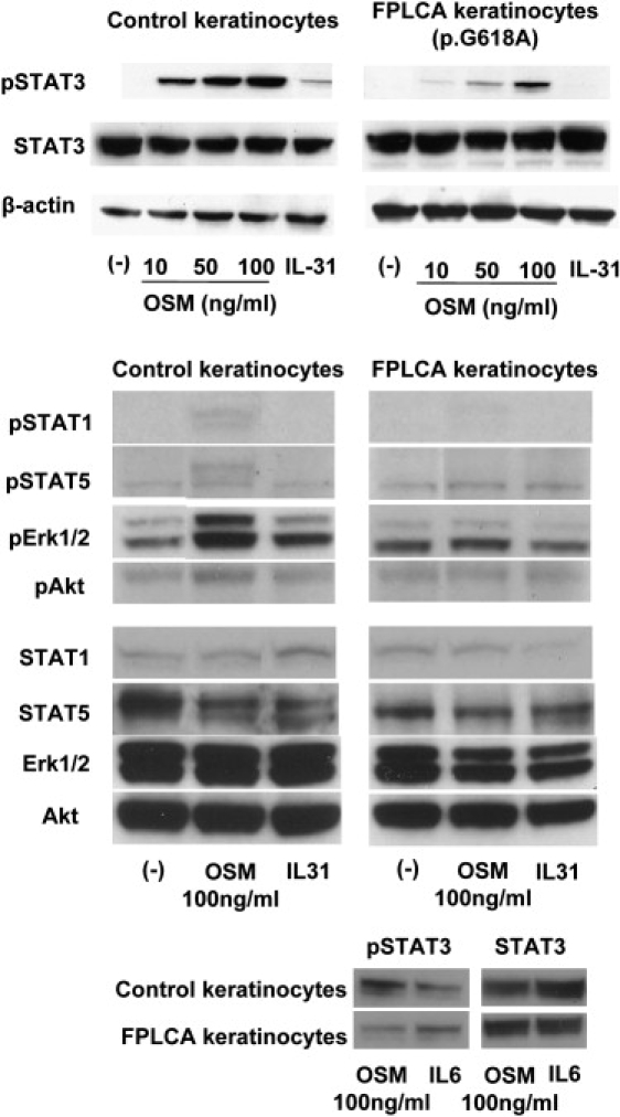
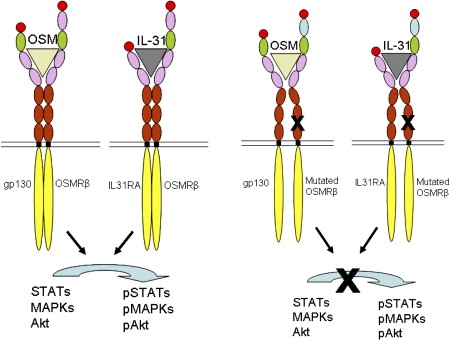
Familial primary localized cutaneous amyloidosis (FPLCA) is an autosomal-dominant disorder associated with chronic skin itching and deposition of epidermal keratin filament-associated amyloid material in the dermis. FPLCA has been mapped to 5p13.1-q11.2, and by candidate gene analysis, we identified missense mutations in the OSMR gene, encoding oncostatin M-specific receptor beta (OSMRbeta), in three families. OSMRbeta is a component of the oncostatin M (OSM) type II receptor and the interleukin (IL)-31 receptor, and cultured FPLCA keratinocytes showed reduced activation of Jak/STAT, MAPK, and PI3K/Akt pathways after OSM or IL-31 cytokine stimulation. The pathogenic amino acid substitutions are located within the extracellular fibronectin type III-like (FNIII) domains, regions critical for receptor dimerization and function. OSM and IL-31 signaling have been implicated in keratinocyte cell proliferation, differentiation, apoptosis, and inflammation, but our OSMR data in individuals with FPLCA represent the first human germline mutations in this cytokine receptor complex and provide new insight into mechanisms of skin itching.


Figures

Figure 1
Figure 1
Clinical, Histological, and Genetic Aspects of FPLCA (A) The pedigree of Family 1 showing autosomal-dominant inheritance; filled symbols represent affected individuals. (B) Clinical features of FPLCA. Skin on the lower leg (individual III-16) appears dry, scaly, and thickened with some hyperpigmentation. (C) At higher magnification, there is accentuation of the normal skin creases and focal skin papules (lichenification). (D) Histopathological features of FPLCA. Skin biopsy of lesional skin shows pigmentary incontinence (arrow) and amorphous eosinophilic material (asterisk) in the superficial papillary dermis (hematoxylin and eosin) (scale bar represents 25 μm). (E) These collections of material show bright fluorescent labeling with thioflavin-T (white arrows) (scale bar represents 25 μm).
Figure 2
Figure 2
The Molecular Basis of FPLCA Involves Heterozygous Missense Mutations in the OSMR Gene (A) DNA sequencing of OSMR. Sequencing of genomic DNA in individuals with FPLCA reveals heterozygous single-nucleotide substitutions in OSMR c.2072T→C in Family 1 and c.1853G→C in Families 2/3 (NM_003999). These convert an isoleucine to threonine and a glycine to alanine, designated p.I691T and p.G618A, respectively. (B) Structural model of OSMRβ depicting the functional domains and the sites of the missense mutations. FNIII, fibronectin type III-like domain; CBD, cytokine binding domain; Ig-like domain, immunoglobulin-like domain. Amino acid (AA) numbers of the 979 amino acid OSMRβ protein are shown on the left of the figure. The missense mutations p.I691T and p.G618A are located within the extracellular FNIII domains. (C) Protein homology and amino acid identity indicate that amino acids p.I691 and p.G618 are well conserved across several species. The numbers at the start of the depicted sequences indicate the amino acid numbers. Underlined amino acids are conserved in all species shown here; p.I691 and p.G618 are highlighted in red.
Figure 3
Figure 3
Schematic Model of IL-6 Family Cytokine Receptors The IL-6 receptor comprises two gp130 and one IL-6R molecules. There are two types of OSM receptors: type I is composed of gp130 and LIFR, whereas type II comprises gp130 and OSMRβ. The IL-31 receptor is composed of OSMRβ and IL31RA. Each domain of the receptors is colored similarly to the schematic shown in Figure 2B.
Figure 4
Figure 4
Western Blotting of Phosphorylated (p)-STATs, pErk1/2, and pAkt of Cultured Keratinocytes Stimulated with OSM or IL-31 Phosphorylation of STATs, Erk1/2, and Akt was observed after stimulation by OSM and IL-31 in normal human keratinocytes, but in FPLCA keratinocytes (here illustrated for Family 2), there was reduced phosphorylation of STATs, Erk1/2, and Akt by OSM and no activation by IL-31 (top and middle). The reduction of pSTAT3 was more clearly demonstrated at lower OSM concentrations (top). Stimulation by IL-6 did not result in altered phosphorylation in FPLCA keratinocytes (bottom).
Figure 5
Figure 5
Structural Representation of OSMR Type II and IL-31 Receptor Signaling in Normal and FPLCA Keratinocytes Each domain of the receptors is colored similarly to the schematic shown in Figure 2B. After binding of OSM or IL-31 to these receptors, the FNIII domains of both receptor subunits interact and signal transduction occurs. The missense mutations seen in our cases occur within the FNIII domains (black crosses), regions critical for receptor dimerization and subsequent signaling. Failure to form conformationally functional receptor complexes in FPLCA leads to reduced receptor signaling after stimulation with either OSM or IL-31.

References

    1. Paus R., Schmelz M., Biro T., Steinhoff M. Frontiers in pruritus research: scratching the brain for more effective itch therapy. J. Clin. Invest. 2006;116:1174–1186. - PMC - PubMed
    1. Breathnach S.M. Amyloid and the amyloidosis of the skin. In: Burns T., Breathnach S., Cox C., Griffiths C., editors. Rook's Textbook of Dermatology. Volume 3. Blackwell Scientific; Oxford: 2004. pp. 57.36–57.51.
    1. Kumakiri M., Hashimoto K. Histogenesis of primary localized cutaneous amyloidosis: sequential change of epidermal keratinocytes to amyloid via filamentous degeneration. J. Invest. Dermatol. 1979;73:150–162. - PubMed
    1. Kobayashi H., Hashimoto K. Amyloidogenesis in organ-limited cutaneous amyloidosis: an antigenic identity between epidermal keratin and skin amyloid. J. Invest. Dermatol. 1983;80:66–72. - PubMed
    1. Schepis C., Siragusa M., Gagliardi M.E., Torre V., Cicciarello R., Albiero F., Cavallari V. Primary macular amyloidosis: an ultrastructural approach to diagnosis. Ultrastruct. Pathol. 1999;23:279–284. - PubMed

Publication types

MeSH terms

Substances