. 2008 Mar 14;283(11):7261-70.
doi: 10.1074/jbc.M702881200. Epub 2008 Jan 7.

A novel role of vascular endothelial cadherin in modulating c-Src activation and downstream signaling of vascular endothelial growth factor

Affiliations

A novel role of vascular endothelial cadherin in modulating c-Src activation and downstream signaling of vascular endothelial growth factor

Chang Hoon Ha et al. J Biol Chem. .

Abstract

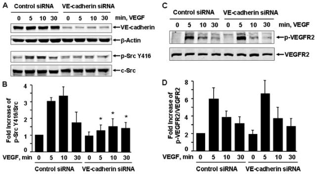
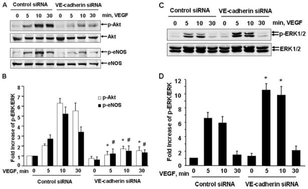
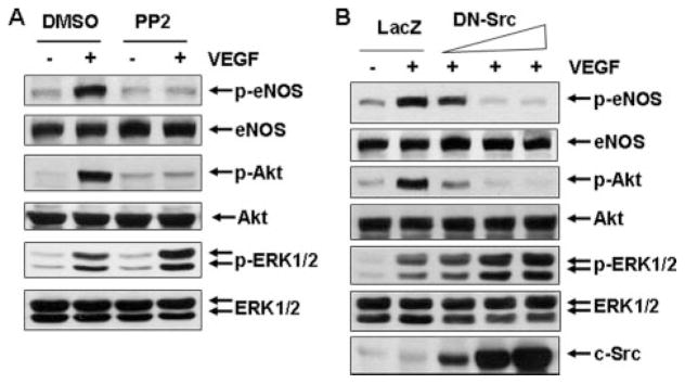
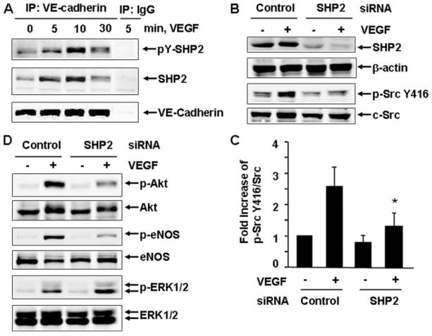
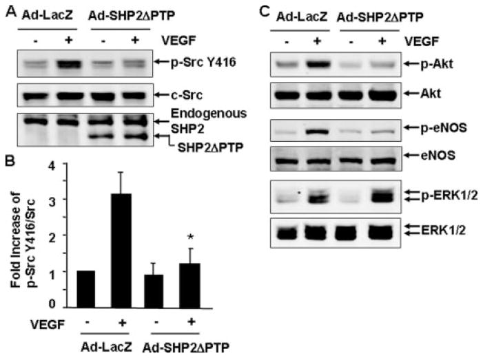
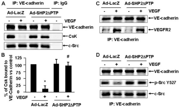
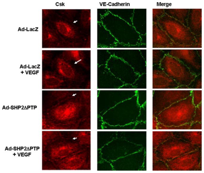
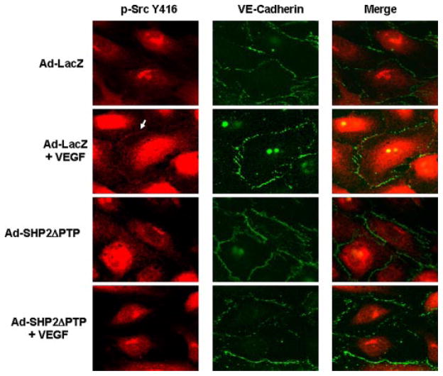
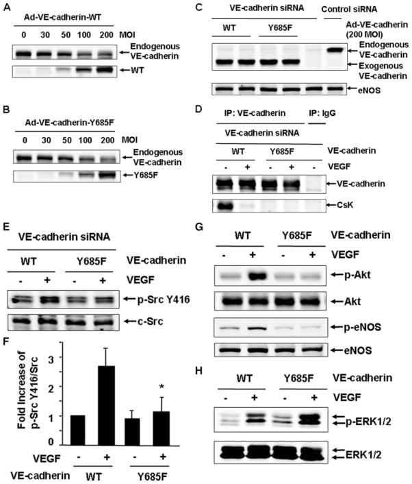
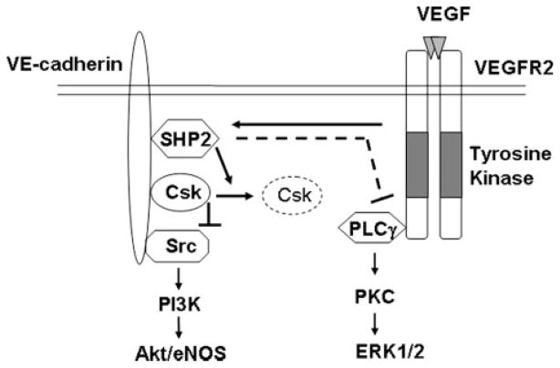
Vascular endothelial growth factor (VEGF) is a potent mediator of angiogenesis and vascular permeability, in which c-Src tyrosine kinase plays an essential role. However, the mechanisms by which VEGF stimulates c-Src activation have remained unclear. Here, we demonstrate that vascular endothelial cadherin (VE-cadherin) plays a critical role in regulating c-Src activation in response to VEGF. In vascular endothelial cells, VE-cadherin was basally associated with c-Src and Csk (C-terminal Src kinase), a negative regulator of Src activation. VEGF stimulated Csk release from VE-cadherin by recruiting the protein tyrosine phosphatase SHP2 to VE-cadherin signaling complex, leading to an increase in c-Src activation. Silencing VE-cadherin with small interference RNA significantly reduced VEGF-stimulated c-Src activation. Disrupting the association of VE-cadherin and Csk through the reconstitution of Csk binding-defective mutant of VE-cadherin also diminished Src activation. Moreover, inhibiting SHP2 by small interference RNA and adenovirus-mediated expression of a catalytically inactive mutant of SHP2 attenuated c-Src activation by blocking the disassociation of Csk from VE-cadherin. Furthermore, VE-cadherin and SHP2 differentially regulates VEGF downstream signaling. The inhibition of c-Src, VE-cadherin, and SHP2 diminished VEGF-mediated activation of Akt and endothelial nitric-oxide synthase. In contrast, inhibiting VE-cadherin and SHP2 enhanced ERK1/2 activation in response to VEGF. These findings reveal a novel role for VE-cadherin in modulating c-Src activation in VEGF signaling, thus providing new insights into the importance of VE-cadherin in VEGF signaling and vascular function.


Figures

FIGURE 1
FIGURE 1. VE-cadherin siRNA inhibited VEGF-induced c-Src activation in endothelial cells
HUVECs were pre-treated with control siRNA or human VE-cadherin siRNA (100 nM) for 48 h and then exposed to VEGF (20 ng/ml) for various times, as indicated. c-Src Tyr-416 phosphorylation in cell lysates was analyzed by Western blot analysis using phosphospecific antibodies (A and B), which recognize c-Src phosphorylated at Tyr-416 (p-Src Y416), and phosphospecific VEGFR2 (p-VEGFR2) (C and D). Total levels of VE-cadherin, β-actin, c-Src, and VEGFR2 were determined by Western blot using their antibodies. Representative immunoblots are shown (A). Quantitative data of Src Tyr-416 phosphorylation and VEGFR2 tyrosine phosphorylation are shown (B) (n = 4).
FIGURE 2
FIGURE 2. VE-cadherin siRNA inhibited VEGF-induced Akt and eNOS activation but promoted ERK1/2 activation
HUVECs were pretreated with control siRNA or human VE-cadherin siRNA (100 nM) for 48 h and then exposed to VEGF (20 ng/ml) for various times, as indicated. The phosphorylation of Akt (Ser-473), eNOS (Ser-1177), and ERK1/2 in cell lysates was determined by Western blot analysis using their phospho-specific antibodies as described under “Experimental Procedures.” Total levels of Akt, eNOS, and ERK1/2 were determined by Western blot using their antibodies. Representative immunoblots (A and C) and quantitative data of protein phosphorylation are shown (B and D) (n = 4).
FIGURE 3
FIGURE 3. Src kinase is required for mediating VEGF-induced Akt and eNOS activation
A, BAECs were pretreated with Src kinase inhibitor PP2 (10 μM) and then stimulated with VEGF (20 ng/ml) for 10 min. DMSO, Me2SO. B, HUVECs were infected with adenovirus encoding LacZ (control, 300 MOI) and adenovirus encoding c-Src-DN at the different doses (30, 100, and 300 MOI) for 24 h and then exposed to VEGF for 10 min. The phosphorylation of Akt, eNOS, and ERK1/2 in cell lysates was determined by Western blot analysis as described under “Experimental Procedures.” Total levels of Akt, eNOS, ERK1/2, and c-Src were determined by Western blot using their antibodies. Representative immunoblots were shown (n = 3).
FIGURE 4
FIGURE 4. SHP2 is essential for mediating VEGF-induced c-Src activation and its downstream signaling
A, BAECs were stimulated with VEGF (20 ng/ml) for the indicated times. The cell lysates were subjected to immunoprecipitation (IP) with VE-cadherin antibodies or mouse IgG as a control followed by immunoblotting with 4G10, SHP2, and VE-cadherin. Representative immunoblots were shown (n = 3). B–D, HUVECs were pretreated with control siRNA and human SHP2 siRNA (150 nM) for 48 h and then exposed to VEGF for 10 min. The phosphorylation of Src Tyr-416, Akt, eNOS, and ERK1/2 in cell lysates was determined by Western blot analysis as described under “Experimental Procedures.” Total levels of SHP2, β-actin, c-Src, Akt, eNOS, and ERK1/2 were determined by Western blot using their antibodies. Representative immunoblots (B and C) and quantitative data of protein phosphorylation were shown (D) (n = 4).
FIGURE 5
FIGURE 5. SHP2 activity is involved in mediating VEGF-induced c-Src activation and its downstream signaling
BAECs were infected with Ad-LacZ (control) or Ad-SHP2ΔPTP for 24 h and then exposed to VEGF (20 ng/ml) for 10 min. The phosphorylation of Src Tyr-416, Akt, eNOS, and ERK1/2 in cell lysates was determined by Western blot analysis as described under “Experimental Procedures.” Total levels of SHP2ΔPTP, c-Src, Akt, eNOS, and ERK1/2 were determined by Western blot using their antibodies. Representative immunoblots (A and C) and quantitative data of protein phosphorylation were shown (B) (n = 5).
FIGURE 6
FIGURE 6. SHP2 regulates c-Src activation by regulating the dissociation of Csk from VE-cadherin in response to VEGF
BAECs were infected with Ad-LacZ (control) or Ad-SHP2ΔPTP for 24 h and then exposed to VEGF (20 ng/ml) for 10 min. The cell lysates were subjected to immunoprecipitation (IP) with VE-cadherin antibodies or mouse IgG as a control followed with immunoblotting with Csk (A), VE-cadherin (A–D), c-Src (A and D), VEGFR2 (C), and phospho-Src Tyr-527 (D). Representative immunoblots (A, C, and D) and quantitative data for the association of Csk and VE-cadherin were shown (B) (n = 6).
FIGURE 7
FIGURE 7. SHP2 regulates Csk dissociation from VE-cadherin in living endothelial cells in response to VEGF
HUVECs were infected with Ad-LacZ (control) or Ad-SHP2ΔPTP for 24 h and then exposed to VEGF (20 ng/ml) for 10 min. The cells were fixed, and Csk and VE-cadherin distribution in cells was analyzed by immunocytochemistry with Csk and VE-cadherin antibodies and by fluorescence microscopy as described under “Experimental Procedures.” Representative images (magnification, ×60) were from four independent experiments.
FIGURE 8
FIGURE 8. SHP2 regulates VEGF-induced c-Src activation at the areas of cell-cell contacts in living endothelial cells
HUVECs were infected with Ad-LacZ (control) or Ad-SHP2ΔPTP for 24 h and then exposed to VEGF (20 ng/ml) for 10 min. The cells were fixed and c-Src Tyr-416 phosphorylation and VE-cadherin distribution in cells was analyzed by immunocytochemistry with phospho-specific c-Src Tyr-416 antibody and VE-cadherin antibody and by fluorescence microscopy as described under “Experimental Procedures.” Representative images (magnification, ×60) were from four independent experiments.
FIGURE 9
FIGURE 9. Csk binding site Tyr-685 in VE-cadherin is critical for mediating c-Src activation in response to VEGF
A and B, HUVECs were infected with adenoviruses encoding mouse VE-cadherin-WT or Y685F at different doses. The endogenous human VE-cadherin and exogenous mouse VE-cadherin were analyzed by immunoblotting. C–H, HUVECs were transfected with VE-cadherin siRNA and then reconstituted with adenoviruses carrying VE-cadherin-WT or Y685F (200 MOI), followed by the exposure of the cells to VEGF (20 ng/ml) for 10 min. The cell lysates were analyzed by immunoblotting with VE-cadherin and eNOS (C) or subjected to immunoprecipitation (IP) with VE-cadherin antibodies or mouse IgG as a control, followed by immunoblotting with Csk and VE-cadherin (D), or analyzed by immunoblotting with the antibodies of phospho-Src Tyr-416 and c-Src (E and F), with the antibodies of phospho-Akt, phospho-eNOS (G), and phospho-ERK1/2 (H). Representative immunoblots and quantitative data for c-Src activation (F) were shown (n = 3).
FIGURE 10
FIGURE 10. Model for a role of VE-cadherin for VEGF-induced c-Src activation and downstream signaling
In resting cells, Csk binds to VE-cadherin through constitutive phosphotyrosine Tyr-685-dependent interaction. c-Src is also associated with VE-cadherin. Such proximate localization of Csk and c-Src in cell adherens junction areas allows Csk to effectively phosphorylate c-Src Tyr-527 and inhibit c-Src activation. When VEGFR2 is activated by VEGF, the VEGF receptor tyrosine kinase phosphorylates VE-cadherin, which permits VE-cadherin to recruit SHP2. The VE-cadherin-bound SHP2 induces release of Csk from VE-cadherin through potentially dephosphorylating VE-cadherin on Csk binding site Tyr-685. Consequently, the c-Src Tyr-527 phosphorylation level decreases, and the c-Src Tyr-416 phosphorylation level increases. The VE-cadherin/SHP2/c-Src module mediates the VEGF-induced phosphatidylinositol 3′-OH-kinase-dependent Akt/eNOS pathway but negatively regulates PLCγ/protein kinase C-dependent ERK activation.

References

    1. Folkman J. Nat Med. 1995;1:27–31. - PubMed
    1. Ferrara N, Gerber HP, LeCouter J. Nat Med. 2003;9:669–676. - PubMed
    1. Carmeliet P. Nat Med. 2003;9:653–660. - PubMed
    1. Weis SM, Cheresh DA. Nature. 2005;437:497–504. - PubMed
    1. Takahashi T, Yamaguchi S, Chida K, Shibuya M. EMBO J. 2001;20:2768–2778. - PMC - PubMed

Publication types

MeSH terms