. 2008 Jan;196(1):149-58.
doi: 10.1677/JOE-07-0391.

Evidence of a possible role for Lys-gamma3-MSH in the regulation of adipocyte function

Affiliations

Evidence of a possible role for Lys-gamma3-MSH in the regulation of adipocyte function

Stephen C Harmer et al. J Endocrinol. 2008 Jan.

Abstract

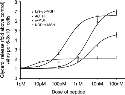
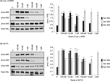
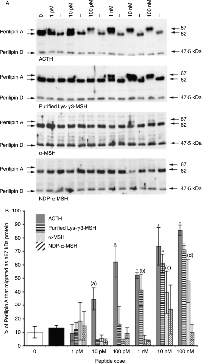
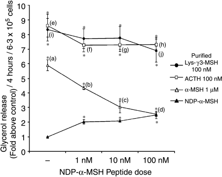
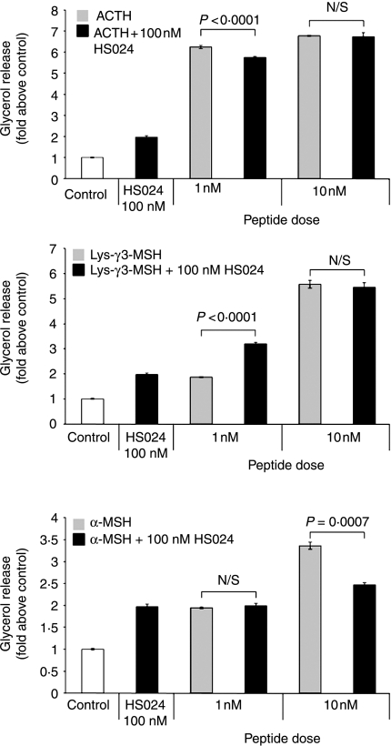
Lys-gamma3-MSH is a melanocortin peptide derived from the C-terminal of the 16 kDa fragment of POMC. The physiological role of Lys-gamma3-MSH is unclear, although it has previously been shown that, although not directly steroidogenic, it can act to potentiate the steroidogenic response of adrenal cortical cells to ACTH. This synergistic effect appears to be correlated with an ability to increase the activity of hormone sensitive lipase (HSL) and therefore the rate of cholesterol ester hydrolysis. Ligand binding studies have suggested that high-affinity binding sites for Lys-gamma3-MSH exist in the adrenal gland and a number of other rat tissues that express HSL, including adipose, skeletal muscle and testes. To investigate the hypothesis that Lys-gamma3-MSH may play a wider role in cholesterol and lipid metabolism, we tested the effect of Lys-gamma3-MSH on lipolysis, an HSL-mediated process, in 3T3-L1 adipocytes. In comparison with other melanocortin peptides, Lys-gamma3-MSH was found to be a potent stimulator of lipolysis. It was also able to phosphorylate HSL at key serine residues and stimulate the hyperphosphorylation of perilipin A. The receptor through which the lipolytic actions of Lys-gamma3-MSH are being mediated is not clear. Attempts to characterise this receptor suggest that either the pharmacology of the melanocortin receptor 5 in 3T3-L1 adipocytes is different from that described when expressed in heterologous systems or the possibility that a further, as yet uncharacterised, receptor exists.


Figures

Figure 1
Figure 1
Lys-γ3-MSH, ACTH, α-MSH and NDP-α-MSH are able to increase lipolysis in 3T3-L1 adipocytes. Results are presented as glycerol release (fold above control), per 4 h, per 6·3×105 cells. Each data point is the mean of the number of responses tested and the error bars represent mean±s.e.m. Curves were fitted using GraphPad Prism and used to calculate EC50 and BMax values for each peptide.
Figure 2
Figure 2
Representative immunoblots showing the effects of (A) Lys-γ3-MSH and (B) ACTH on the phosphorylation state of HSL at specific serine residues in 3T3-L1 adipocytes. The phosphorylation state of HSL was determined, after a 20-min stimulation in the presence or absence of Lys-γ3-MSH or ACTH at increasing dose from 1 pM to 100 nM, by analysing the degree of phosphorylation of serine residues 563, 565 and 660 using antibodies specific for each phosphorylated serine site. (0), no peptide added. Densitometric analysis was performed on the results of three independent experiments and is presented as the means of the phosphorylated HSL content divided by the mean of the total HSL content for the same sample. The error bars represent the mean±s.e.m. *P<0·05 and **P<0·01 indicate a significant difference from the corresponding untreated value.
Figure 3
Figure 3
(A) Analysis of the effect of Lys-γ3-MSH, ACTH, α-MSH and NDP-α-MSH on the phosphorylation state of perilipin A in 3T3-L1 adipocytes. The phosphorylation state of perilipin A was determined by analysing the migratory rate of perilipin A, on 6·5% SDS-PAGE gels, either 62 kDa (unstimulated) or 67 kDa (stimulated, hyperphosphorylated). (−), no peptide added. (0), time point zero. (B) Densitometric analysis of the effect of Lys-γ3-MSH, ACTH, α-MSH and NDP-α-MSH on the phosphorylation state of perilipin A in 3T3-L1 adipocytes. The phosphorylation state of perilipin A was determined by analysing the migratory rate of perilipin A, on 6·5% SDS-PAGE gels, either 62 kDa (unstimulated) or 67 kDa (stimulated, hyperphosphorylated). All results presented, except those at time point 0 (0) and (−) (no peptide added), are the average of three responses from independent experiments. Results presented at time point (0) and (−) are the average of 12 responses from three independent experiments. The error bars represent the mean ±s.e.m. (a) (P=0·0194) is significantly different from the control (−) value. (b) (P=0·0022) is significantly different from the control (−) value. (c) (P=0·0061) is significantly different from the control (−) value. (d) (P=0·004) is significantly different from the control (−) value. *(P<0·0001) indicates that values are significantly different from the control (−) value.
Figure 4
Figure 4
The potent melanocortin receptor agonist NDP-α-MSH can reduce the lipolytic activity of α-MSH but not ACTH or Lys-γ3-MSH in 3T3-L1 adipocytes when compared with the lipolytic activity seen in response to NDP-α-MSH alone. The lipolytic activity of ACTH, Lys-γ3-MSH and α-MSH were determined in the presence or absence of an increasing dose of NDP-α-MSH. Results are presented as glycerol release (fold above control), per 4-h incubation, per 6·3×105 cells. The error bars represent mean±s.e.m. *(P<0·0001) significant difference from the control level. (b), (c) and (d) are (P<0·0001) significantly different from (a). (f), (g) and (h) are (P<0·05) significantly different from (e). (j) is (P<0·05) significantly different from (i).
Figure 5
Figure 5
The MC1, MC3, MC4 and MC5 receptor antagonist HS024 can reduce the lipolytic activity of α-MSH, but increases the lipolytic response to Lys-γ3-MSH in 3T3-L1 adipocytes when compared with the lipolytic activity seen in response to HS024 alone. The lipolytic activity of two sub-maximal doses of ACTH, Lys-γ3-MSH and α-MSH were determined in the presence or 100 nM HS024. Results are presented as glycerol release (fold above control), per 4-h incubation, per 6·3×105 cells. The error bars represent mean±s.e.m. All treatments resulted in a significant (P<0·0001) increase in lipolysis when compared with control cells. Between-treatment differences (with their respective P values) are shown on the figure.

References

    1. Al-Dujaili EA, Hope J, Estivariz FE, Lowry PJ, Edwards CR. Circulating human pituitary pro-gamma-melanotropin enhances the adrenal response to ACTH. Nature. 1981;29:156–159. - PubMed
    1. Anthonsen MW, Rönnstrand L, Wernstedt C, Degerman E, Holm C. Identification of novel phosphorylation sites in hormone-sensitive lipase that are phosphorylated in response isoproterenol and govern activation properties in vitro. Journal of Biological Chemistry. 1998;273:215–221. - PubMed
    1. Bennett HPJ. Use of ion-exchange Sep-Pak cartridges in the batch fractionation of pituitary peptides. Journal of Chromatography. 1986;359:383–390. - PubMed
    1. Bicknell AB, Lomthaisong K, Woods RJ, Hutchinson EG, Bennett HP, Gladwell RT, Lowry PJ. Characterisation of a serine protease that cleaves pro-gamma-melanotropin at the adrenal to stimulate growth. Cell. 2001;105:903–912. - PubMed
    1. Bohlen P, Esch F, Shibasaki T, Baird A, Ling N, Guillemin R. Isolation and characterization of γ1-melanotropin-like peptide from bovine neurointermediate pituitary. FEBS Letters. 1981;128:67–70. - PubMed

Publication types

MeSH terms