. 2008 Mar 1;111(5):2674-80.
doi: 10.1182/blood-2007-08-110205. Epub 2008 Jan 7.

Semaphorin 3A suppresses VEGF-mediated angiogenesis yet acts as a vascular permeability factor

Affiliations

Semaphorin 3A suppresses VEGF-mediated angiogenesis yet acts as a vascular permeability factor

Lisette M Acevedo et al. Blood. .

Abstract

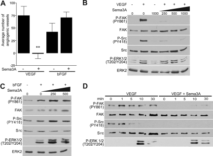
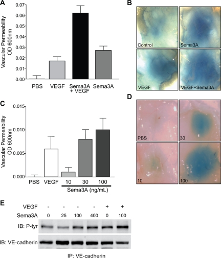
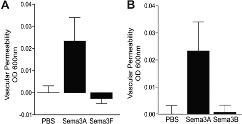
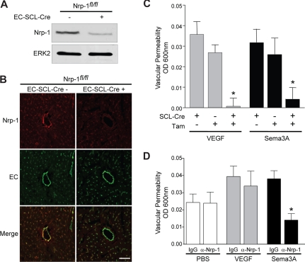
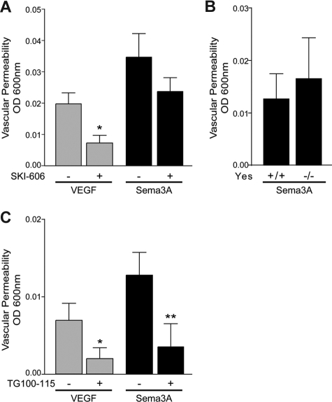
Semaphorin 3A (Sema3A), a known inhibitor of axonal sprouting, also alters vascular patterning. Here we show that Sema3A selectively interferes with VEGF- but not bFGF-induced angiogenesis in vivo. Consistent with this, Sema3A disrupted VEGF- but not bFGF-mediated endothelial cell signaling to FAK and Src, key mediators of integrin and growth factor signaling; however, signaling to ERK by either growth factor was unperturbed. Since VEGF is also a vascular permeability (VP) factor, we examined the role of Sema3A on VEGF-mediated VP in mice. Surprisingly, Sema3A not only stimulated VEGF-mediated VP but also potently induced VP in the absence of VEGF. Sema3A-mediated VP was inhibited either in adult mice expressing a conditional deletion of endothelial neuropilin-1 (Nrp-1) or in wild-type mice systemically treated with a function-blocking Nrp-1 antibody. While both Sema3A- and VEGF-induced VP was Nrp-1 dependent, they use distinct downstream effectors since VEGF- but not Sema3A-induced VP required Src kinase signaling. These findings define a novel role for Sema3A both as a selective inhibitor of VEGF-mediated angiogenesis and a potent inducer of VP.


Figures

Figure 1
Figure 1
Sema3A interferes with VEGF- but not bFGF-induced angiogenesis. (A) Inhibition of VEGF- but not bFGF-mediated angiogenesis by Sema3A was determined by treating CAMs with PBS, VEGF (40 μg/mL), and bFGF (40 μg/mL) with or without Sema3A (50 μg/mL) for 3 days (n = 15, **P < .01). Error bars represent SEM. (B,C) Increasing concentrations of Sema3A (ng/mL) selectively suppresses FAK and Src activation in VEGF- but not bFGF-stimulated (5 minutes) cells. Phosphorylation of FAK, Src, and ERK was determined through immunoblotting. (D) HUVECs were treated with 250 ng/mL Sema3A prior to stimulation with VEGF throughout various time points. These experiments were repeated multiple times with similar results.
Figure 2
Figure 2
Sema3A potentiates VEGF-induced permeability and induces VP on its own. (A) Sema3A potentiation of VEGF-mediated VP was determined by subcutaneous injection of PBS, VEGF (40 μg/mL), Sema3A (30 μg/mL), or VEGF with Sema3A in Balb/c mice. Evans blue dye (1.5%) extravasation was quantitated (n = 8). Data represent means plus or minus SEM. (B) Representative images of Sema3A- and/or VEGF-induced permeability. (C) Sema3A dose-dependently induces VP as shown by injecting mice with increasing amounts of Sema3A (μg/mL), VEGF, or PBS as control (n = 6). Evans blue dye (1%) extravasation was quantitated. Data represent mean plus or minus SEM. This experiment was repeated twice with similar results. (D) Representative images of dose-dependent Sema3A-induced permeability. (E) Sema3A dose-dependently induces tyrosine phosphorylation of VE-cadherin as demonstrated by treatment of HUVECs with increasing amounts of Sema3A (ng/mL), VEGF (50 ng/mL), or both. This experiment was repeated multiple times with similar results.
Figure 3
Figure 3
Unlike Sema3A, Sema3F and Sema3B do not induce VP. (A) Balb/c mice were injected with 30 μg/mL Sema3A or Sema3F (n = 9), and 1% Evans blue dye extravasation was quantitated. Data represent mean plus or minus SEM. (B) Balb/c mice were injected with 30 μg/mL Sema3A or Sema3B (n = 6). Data represent means plus or minus SEM.
Figure 4
Figure 4
Sema3A and VEGF require Nrp-1 expression to induce vascular permeability. (A) To determine knockdown of Nrp-1 expression in inducible, endothelial-specific knockouts, mice were injected with 20 mg/mL tamoxifen (Tam) for 2 weeks to excise the floxed exon. Heart lysates from EC-SCL-CreERT–positive or –negative Nrp-1fl/fl mice were immunoblotted with antibodies against Nrp-1 or ERK2 as a loading control. (B) Cryosections (5 μm) from Tam-treated EC-SCL-CreERT–positive or –negative Nrp-1fl/fl hearts were stained with antibodies against Nrp-1 (red) and the endothelial markers (EC, green). Confocal images were taken at 400× magnification (scale bar represents 50 μm). (C) Knockdown of Nrp-1 expression disrupts Sema3A- and VEGF-mediated VP. Mice treated with Tam or vehicle were injected subcutaneously with Sema3A, VEGF, or PBS. Dye extravasation was quantitated (n = 8, *P < .05). (D) A function-blocking monoclonal antibody against Nrp-1 inhibits Sema3A- but not VEGF-induced VP. Balb/c mice were injected with an anti–Nrp-1 or IgG (50 μg/mL intravenously) 30 minutes prior to injection of VEGF, Sema3A, or PBS (n = 9, *P < .05). Error bars represent SEM.
Figure 5
Figure 5
Induction of permeability by Sema3A is Src independent, but is blocked by PI3Kγ/δ inhibition. (A) Treatment with the Src family kinase inhibitor SKI-606 (5 mg/kg intraperitoneally) prior to injection with Sema3A, VEGF, or PBS significantly inhibits VEGF- but not Sema3A-mediated VP (n = 10, *P < .05). (B) Yes+/+ and Yes−/− mice show similar VP response when injected with Sema3A or PBS (n = 8). (C) Sema3A- and VEGF-induced VP depends on PI3Kγ/δ as determined by treatment of mice with the PI3Kγ/δ inhibitor TG100–115 (5 mg/kg intraperitoneally) prior to injection with Sema3A, VEGF, or PBS (n = 5, *P < .05, **P < .01). Error bars represent SEM.

References

    1. Yazdani U, Terman JR. The semaphorins. Genome Biol. 2006 7:211. [Accessed May 23, 2006]; ( http://genomebiology.com) - PMC - PubMed
    1. Serini G, Valdembri D, Zanivan S, et al. Class 3 semaphorins control vascular morphogenesis by inhibiting integrin function. Nature. 2003;424:391–397. - PubMed
    1. Miao HQ, Soker S, Feiner L, Alonso JL, Raper JA, Klagsbrun M. Neuropilin-1 mediates collapsin-1/semaphorin III inhibition of endothelial cell motility: functional competition of collapsin-1 and vascular endothelial growth factor-165. J Cell Biol. 1999;146:233–242. - PMC - PubMed
    1. Shoji W, Isogai S, Sato-Maeda M, Obinata M, Kuwada JY. Semaphorin3a1 regulates angioblast migration and vascular development in zebrafish embryos. Development. 2003;130:3227–3236. - PubMed
    1. Torres-Vazquez J, Gitler AD, Fraser SD, et al. Semaphorin-plexin signaling guides patterning of the developing vasculature. Dev Cell. 2004;7:117–123. - PubMed

Publication types

MeSH terms

Substances