. 2008 Jan 15;105(2):710-5.
doi: 10.1073/pnas.0708110105. Epub 2008 Jan 7.

Role for lysosomal enzyme beta-hexosaminidase in the control of mycobacteria infection

Affiliations

Role for lysosomal enzyme beta-hexosaminidase in the control of mycobacteria infection

Ingrid Chou Koo et al. Proc Natl Acad Sci U S A. .

Abstract

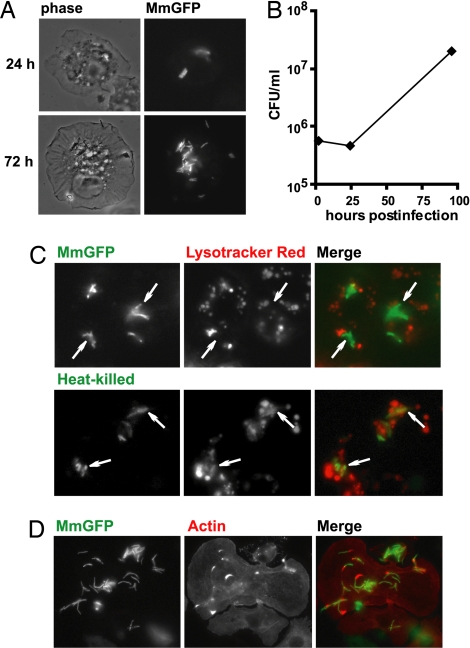
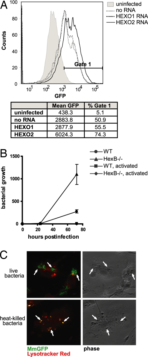
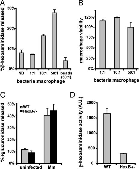
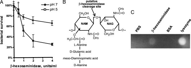
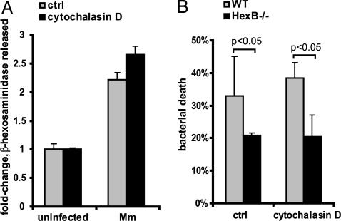
The pathogenic mycobacteria that cause tuberculosis (TB) and TB-like diseases in humans and animals elude sterilizing immunity by residing within an intracellular niche in host macrophages, where they are protected from microbicidal attack. Recent studies have emphasized microbial mechanisms for evasion of host defense; less is known about mycobactericidal mechanisms that remain intact during initial infection. To better understand macrophage mechanisms for restricting mycobacteria growth, we examined Mycobacterium marinum infection of Drosophila S2 cells. Among approximately 1,000 host genes examined by RNAi depletion, the lysosomal enzyme beta-hexosaminidase was identified as an important factor in the control of mycobacterial infection. The importance of beta-hexosaminidase for restricting mycobacterial growth during mammalian infections was confirmed in macrophages from beta-hexosaminidase knockout mice. Beta-hexosaminidase was characterized as a peptidoglycan hydrolase that surprisingly exerts its mycobactericidal effect at the macrophage plasma membrane during mycobacteria-induced secretion of lysosomes. Thus, secretion of lysosomal enzymes is a mycobactericidal mechanism that may have a more general role in host defense.


Conflict of interest statement

The authors declare no conflict of interest.

Figures

Fig. 1.
Fig. 1.
Mm infects and proliferates in S2 cells. (A) S2 cells were infected by MmGFP and visualized 24 and 72 h after infection. Phase-contrast images are shown on the left, and immunofluorescence is shown on the right. (B) Mm growth in S2 cells was enumerated by colony-forming units over a 4-day period. (C) MmGFP-infected S2 cells were stained with Lysotracker red. (Upper) Uptake of live bacilli. (Lower) Uptake of heat-killed bacilli. Fusion of phagosomes containing heat-killed Mm-containing with lysosomes is indicated by overlap of the entire bacterium with red fluorescence, demonstrating the low pH (5.5) of the phagosome. Examples of bacteria are indicated by arrows. (D) Infected S2 cells were fixed and stained with Alexa Fluor 594 phalloidin (red). Actin tails and clouds were found in proximity to intracellular bacteria.
Fig. 2.
Fig. 2.
Effect of β-hexosaminidase on intracellular growth of Mm. (A) Flow cytometry histogram of S2 cells infected by MmGFP 48 h after initiation of infection. The fluorescence of Mm was greater in HEXO2 RNAi-treated S2 cells, despite equivalent initial uptake. Histogram is representative of three independent experiments. (B) Growth curve of Mm in WT or HexB−/− BMDM. Mm shows increased intracellular growth in HexB−/− BMDM. Activated BMDM (by treatment with IFN-γ and LPS) did not support growth of the bacilli. The graph shown is normalized for bacterial uptake (after washing of monolayer) and representative of three independent experiments. (C) MmGFP-infected HexB−/− BMDM were stained with Lysotracker red 24 h after initial bacterial uptake. (Upper) Uptake of live bacilli. (Lower) Uptake of heat-killed bacilli. Fusion of phagosomes containing heat-killed Mm with lysosomes is indicated by overlap of the entire bacterium with red fluorescence, demonstrating the low pH (5.5) of the phagosome. Examples of bacteria are indicated by arrows.
Fig. 3.
Fig. 3.
Mm induces secretion of lysosomes from macrophages. (A) Secretion of β-hexosaminidase by BMDM was measured 2 h after treatment with buffer (NB, no bacteria), Mm (MOI of 1, 10, or 50), or latex beads (beads, 50:1). (B) Viability of BMDM was measured 2 h after addition of inoculum. Cell viability is displayed as Absinfected/Absuninfected. (C) Secretion of lysosomal enzyme β-glucuronidase was measured 2 h after treatment of WT and HexB−/− BMDM with buffer (uninfected) or Mm (Mm). (D) Secretion of β-hexosaminidase was measured in Mm-infected WT or HexB−/− BMDM. β-Hexosaminidase activity is displayed in arbitrary units (A.U.). All assays were done in triplicate.
Fig. 4.
Fig. 4.
β-Hexosaminidase is a mycobactericidal peptidoglycan hydrolase. (A) Mm was incubated with 0, 0.5, 1, 2, and 4 units/ml human β-hexosaminidase. Bacterial survival was enumerated as colony-forming units on 7H10 agar. Assays were done in triplicate, at pH values of 5 and 7. (B) β-Hexosaminidase is predicted to hydrolyze the β-1,4-linked glycosidic bond between NAM and NAG, as shown. (C) PG hydrolase activity of β-hexosaminidase was assessed by zymography. PBS and BSA were negative controls, and lysozyme was used as a positive control.
Fig. 5.
Fig. 5.
β-Hexosaminidase exerts mycobactericidal effects at the cell surface in the absence of phagocytosis. (A) Secretion of β-hexosaminidase was measured in response to Mm (MOI of 50) in the presence or absence of 1 μM cytochalasin D. (B) Mm killing by WT and HexB−/− BMDM was assessed 2 h after initiation of interaction in the presence or absence of cytochalasin D. All assays were done in triplicate.

References

    1. Frieden TR, Sterling TR, Munsiff SS, Watt CJ, Dye C. Lancet. 2003;362:887–899. - PubMed
    1. Stamm LM, Brown EJ. Microbes Infect. 2004;6:1418–1428. - PubMed
    1. Ramakrishnan L, Falkow S. Infect Immun. 1994;62:3222–3229. - PMC - PubMed
    1. Rook GA, Taverne J, Leveton C, Steele J. Immunology. 1987;62:229–234. - PMC - PubMed
    1. Holland SM. Am J Med Sci. 2001;321:49–55. - PubMed

Publication types

Substances

LinkOut - more resources