. 2008 Mar;16(3):466-73.
doi: 10.1038/sj.mt.6300395. Epub 2008 Jan 8.

Freeze-dried tendon allografts as tissue-engineering scaffolds for Gdf5 gene delivery

Affiliations

Freeze-dried tendon allografts as tissue-engineering scaffolds for Gdf5 gene delivery

Patrick Basile et al. Mol Ther. 2008 Mar.

Abstract

Tendon reconstruction using grafts often results in adhesions that limit joint flexion. These adhesions are precipitated by inflammation, fibrosis, and the paucity of tendon differentiation signals during healing. In order to study this problem, we developed a mouse model in which the flexor digitorum longus (FDL) tendon is reconstructed using a live autograft or a freeze-dried allograft, and identified growth and differentiation factor 5 (Gdf5) as a therapeutic target. In this study we have investigated the potential of rAAV-Gdf5 -loaded freeze-dried tendon allografts as "therapeutically endowed" tissue-engineering scaffolds to reduce adhesions. In reporter gene studies we have demonstrated that recombinant adeno-associated virus (rAAV)-loaded tendon allografts mediate efficient transduction of adjacent soft tissues, with expression peaking at 7 days. We have also demonstrated that the rAAV-Gdf5 vector significantly accelerates wound healing in an in vitro fibroblast scratch model and, when loaded onto freeze-dried FDL tendon allografts, improves the metatarsophalangeal (MTP) joint flexion to a significantly greater extent than the rAAV-lacZ controls do. Collectively, our data demonstrate the feasibility and efficacy of therapeutic tendon allograft processing as a novel paradigm in tissue engineering in order to address difficult clinical problems such as tendon adhesions.


Figures

Figure 1
Figure 1
Transduction efficacy of freeze-dried tendon grafts in vitro and in vivo. 3mm freeze-dried mouse FDL tendon allografts (a) were loaded with 5×10⁹ transducing units of rAAV-lacZ, and incubated on a confluent monolayer of HEK 293 cells for 48 hours (arrow). Representative micrographs of x-gal stained cultures show large numbers of LacZ+ cells proximal to the graft (b), and sparse staining in peripheral fields away from the graft (c). rAAV-lacZ loaded FDL allografts were also transplanted into FDL tendon defects of mice (n=4). Representative micrographs of one end of the rAAV-lacZ loaded FDL allografts stained with antibodies against β-galactosidase at 7 days (d) and 14 days (e) post-transplantation. Of note is the lack of viable cells and any staining in the freeze-dried allografts (asterisks) that are surrounded by hypercellular and intensely stained fibrotic tissue. (C) The specificity of the staining was verified by the absence of non-specific staining in negative controls (secondary antibody only – f). S indicates remnants of the repair suture.
Figure 2
Figure 2
Kinetics and biodistribution rAAV-mediated transduction via processed tendon allografts. Temporal bioluminescence images (BLI) of a representative mouse grafted with a freeze-dried FDL tendon allograft loaded with rAAV-Luc over 21 days show the localized biodistribution of rAAV-Luc transduction (heat map-yellow arrows) at the site of allograft implantation in the hind foot (a). Kinetics of in vivo rAAV transduction (b) based on average BLI signal intensity computed from measurements of total integrated light signal (photons emitted/cm2/sec) emitted from a standardized region of interest (ROI) in a standard 3 minute time interval (mean ± SEM; n=4).
Figure 3
Figure 3
Functional verification of the rAAV-Gdf5 vector. HEK293 cells were grown in 6-well plates and transfected with: 1) pUC19, 2) pSPORT-Gdf5, or pAAV-Gdf5, and 48hrs later total RNA was harvested from the cells. The mRNA was reverse transcribed and used as the template for PCR with Gdf5 specific primers. 4) The pSPORT-Gdf5 plasmid was used for template in the positive control. The ethidium bromide stained agarose gel shows the predicted 485bp PCR product (a – Top). HEK293 cells were grown in 6-well plates and infected with the indicated amount of rAAV-lacZ or rAAV-Gdf5 (5.0×10⁷ particles per ml). After 48hrs of culture, the supernatants were collected and 30µl was used for Western blotting with GDF-5 specific antibodies. 10ng of recombinant murine GDF-5 was used as a positive control. Autoradiography of the Western blot reveals the predicted 13.7kDa GDF-5 protein (a – Bottom). Microwound monolayer assay: 80% confluent 3T3 cells were growth arrested for 24 hours then microwounded by passing a pipette tip across the culture well (b) and treated with 0.5% bovine calf serum (BCS) and 5.0×10⁷ particles/ml of either rAAV-lacZ or rAAV-Gdf5. The average width of the defect was digitally measured over time and the wound width normalized to the time zero width [w(t)/w(0)] versus time was plotted(c). Healing time constants (τ) for the different treatments were computed and plotted as mean ± SEM (d). Note that higher τ values indicate slower wound healing rates. In a separate experiment, 3T3 cells grown to 80% confluence were microwounded and treated with 0.5% BCS and incremental doses of rmGDF-5. Data presented are mean ± SEM for the healing time constant (τ) for the different doses of the GDF-5 protein treatments (e). Asterisks indicate significant differences (p<0.01; n=6 per treatment) compared to untreated controls.
Figure 4
Figure 4
rAAV-Gdf5 loading of freeze-dried allografts improves the MTP flexion range of motion and the gliding function of reconstructed FDL tendons while maintaining their biomechanical properties. Mice had their FDL tendons reconstructed with freeze dried allografts loaded with rAAV-Gdf5 (treated) or rAAV-lacZ (controls) and sacrificed at 14 and 28 days post-operatively (n=9 per treatment per time point). The operated hind feet were harvested and subjected to the MTP flexion test to determine the MTP joint flexion ROM (a) and the Gliding Coefficient (b). The tendons were then isolated and tested biomechanically to determine their breaking (maximum) tensile force (c) and their linear tensile stiffness (d). Data presented are mean ± SEM. Asterisks indicate significant differences compared to time-matched controls (p<0.05).
Figure 5
Figure 5
rAAV-Gdf5 loading of freeze-dried allografts mediates de novo GDF-5 protein synthesis by the host cells at the periphery of the implanted allograft. Representative immunohistochemical sections of the rAAV-lacZ (a) or rAAV-Gdf5 (b) loaded FDL tendon allografts at 14 days post grafting and stained with anti-mouse GDF-5 antibody. Of note is the matrix-bound GDF-5 (positive staining indicated by arrows) presumably synthesized by the transduced host cells surrounding the rAAV-Gdf5 treated allografts (asterisk) that is absent in the rAAV-lacZ treated graft.
Figure 6
Figure 6
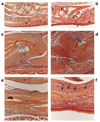
rAAV-Gdf5 loading of freeze-dried allografts mediates cellular repopulation of the graft and remodeling of the fibrotic scar tissue. Representative histological sections of the rAAV-lacZ (a,c,e) or rAAV-Gdf5 (b,d,f) loaded FDL tendon allografts at 14 days post grafting and stained with Alcian Blue and Orange G. Micrographs at 4X (a and b) show the implanted grafts with their anatomical relationships to the surrounding tissue. Boxed regions are shown in the magnified micrographs (20X) that show the distal ends of both grafts (c and d) and the middle segment of the grafts (e and f). Tissues marked by numbers are: 1) Talus, 2) Tarsal bones, 3) Metatarsal bone, 4) FDL tendon allograft, and 5) fibrotic/inflammatory tissue. S indicates remnants of suture. Arrows in f indicate a remodeled tissue that appears to align and integrate with the rAAV-Gdf5 loaded allograft which also seems to have been repopulated with host cells compared to the mostly acellular rAAV-lacZ loaded allograft (e).

Similar articles

Cited by

References

    1. Lilly SI, Messer TM. Complications after treatment of flexor tendon injuries. J Am Acad Orthop Surg. 2006;14:387–396. - PubMed
    1. Gelberman RH, Manske PR. Factors influencing flexor tendon adhesions. Hand Clin. 1985;1:35–42. - PubMed
    1. Silva MJ, Boyer MI, Gelberman RH. Recent progress in flexor tendon healing. J Orthop Sci. 2002;7:508–514. - PubMed
    1. Leddy JP. Flexor Tendons - Acute Injuries. In: Green DP, editor. Operative Hand Surgery. Vol. 3. New York: Churchill Livingstone; 1988. pp. 1935–1968.
    1. Whitlock PW, Smith TL, Poehling GG, Shilt JS, Van Dyke M. A naturally derived, cytocompatible, and architecturally optimized scaffold for tendon and ligament regeneration. Biomaterials. 2007;28:4321–4329. - PubMed

Publication types

MeSH terms

Substances