

Endocytic pathways: combined scanning ion conductance and surface confocal microscopy study
- PMID: 18180951
- PMCID: PMC2270919
- DOI: 10.1007/s00424-007-0410-4
Endocytic pathways: combined scanning ion conductance and surface confocal microscopy study
Abstract
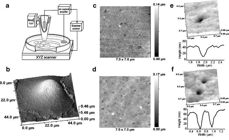
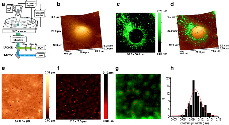
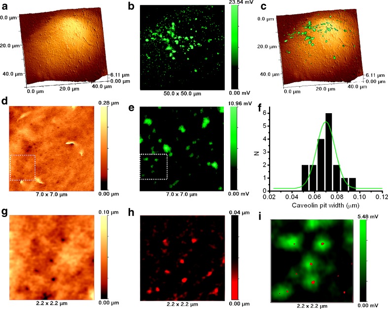
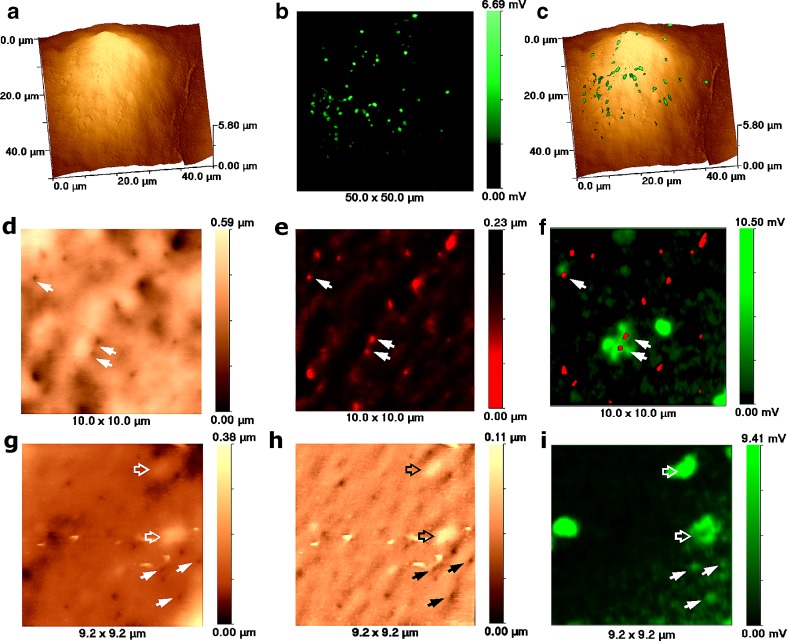
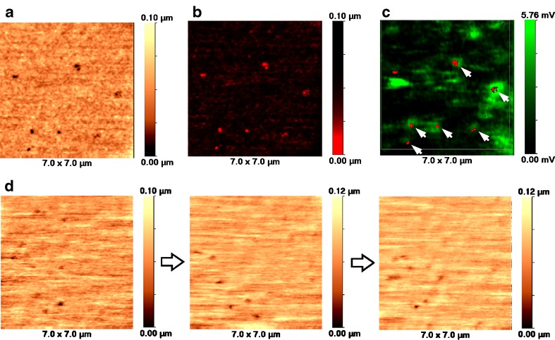
We introduce a novel high resolution scanning surface confocal microscopy technique that enables imaging of endocytic pits in apical membranes of live cells for the first time. The improved topographical resolution of the microscope together with simultaneous fluorescence confocal detection produces pairs of images of cell surfaces sufficient to identify single endocytic pits. Whilst the precise position and size of the pit is detected by the ion conductance microscope, the molecular nature of the pit, e.g. clathrin coated or caveolae, is determined by the corresponding green fluorescent protein fluorescence. Also, for the first time, we showed that flotillin 1 and 2 can be found co-localising with approximately 200-nm indentations in the cell membrane that supports involvement of this protein in endocytosis.
Figures
Similar articles
-
An alternative mechanism of clathrin-coated pit closure revealed by ion conductance microscopy.J Cell Biol. 2012 May 14;197(4):499-508. doi: 10.1083/jcb.201109130. Epub 2012 May 7. J Cell Biol. 2012. PMID: 22564416 Free PMC article.
-
Correlative SICM-FCM reveals changes in morphology and kinetics of endocytic pits induced by disease-associated mutations in dynamin.FASEB J. 2019 Jul;33(7):8504-8518. doi: 10.1096/fj.201802635R. Epub 2019 Apr 24. FASEB J. 2019. PMID: 31017801 Free PMC article.
-
A hybrid scanning mode for fast scanning ion conductance microscopy (SICM) imaging.Ultramicroscopy. 2012 Oct;121(11):1-7. doi: 10.1016/j.ultramic.2012.06.015. Epub 2012 Jul 5. Ultramicroscopy. 2012. PMID: 22902298 Free PMC article.
-
Endocytosis without clathrin coats.Trends Cell Biol. 2001 Oct;11(10):406-12. doi: 10.1016/s0962-8924(01)02107-9. Trends Cell Biol. 2001. PMID: 11567873 Review.
-
Molecular mechanisms of clathrin-independent endocytosis.J Cell Sci. 2009 Jun 1;122(Pt 11):1713-21. doi: 10.1242/jcs.033951. J Cell Sci. 2009. PMID: 19461071 Free PMC article. Review.
Cited by
-
Scanning Ion Conductance Microscopy.Chem Rev. 2021 Oct 13;121(19):11726-11768. doi: 10.1021/acs.chemrev.0c00962. Epub 2020 Dec 9. Chem Rev. 2021. PMID: 33295182 Free PMC article. Review.
-
Multifunctional scanning ion conductance microscopy.Proc Math Phys Eng Sci. 2017 Apr;473(2200):20160889. doi: 10.1098/rspa.2016.0889. Epub 2017 Apr 12. Proc Math Phys Eng Sci. 2017. PMID: 28484332 Free PMC article. Review.
-
Cellular endocytosis and gene delivery.Mol Med. 2010 May-Jun;16(5-6):222-9. doi: 10.2119/molmed.2009.00101. Epub 2010 Feb 3. Mol Med. 2010. PMID: 20454523 Free PMC article. Review.
-
Scanning ion conductance microscopy: a nanotechnology for biological studies in live cells.Front Physiol. 2013 Jan 14;3:483. doi: 10.3389/fphys.2012.00483. eCollection 2012. Front Physiol. 2013. PMID: 23335899 Free PMC article.
-
An alternative mechanism of clathrin-coated pit closure revealed by ion conductance microscopy.J Cell Biol. 2012 May 14;197(4):499-508. doi: 10.1083/jcb.201109130. Epub 2012 May 7. J Cell Biol. 2012. PMID: 22564416 Free PMC article.
References
Publication types
MeSH terms
Substances
Grants and funding
LinkOut - more resources
Full Text Sources
