. 2008 Feb 5;47(5):1381-92.
doi: 10.1021/bi701189c. Epub 2008 Jan 9.

Structural impact of three Parkinsonism-associated missense mutations on human DJ-1

Affiliations

Structural impact of three Parkinsonism-associated missense mutations on human DJ-1

Mahadevan Lakshminarasimhan et al. Biochemistry. .

Abstract

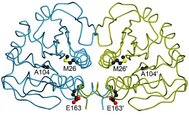
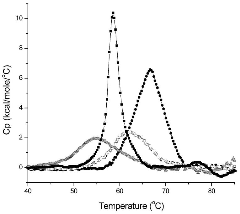
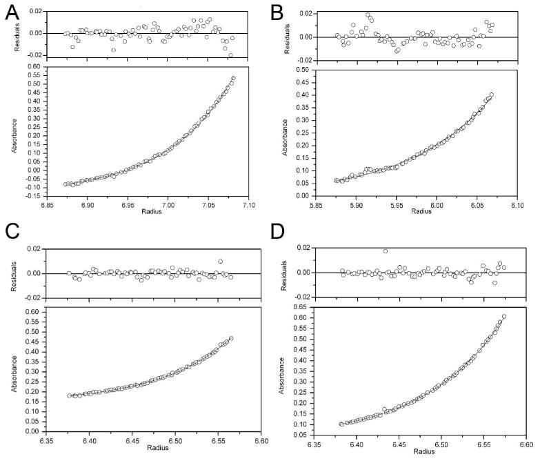
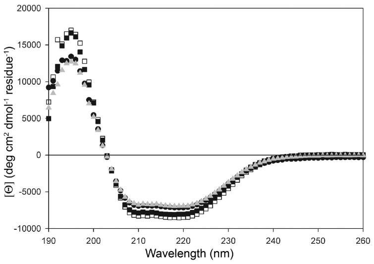
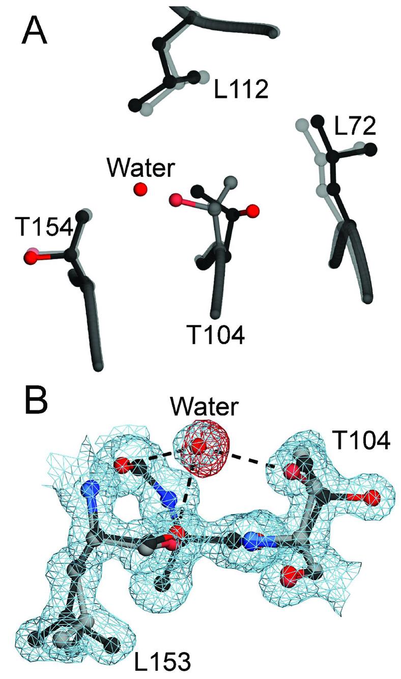
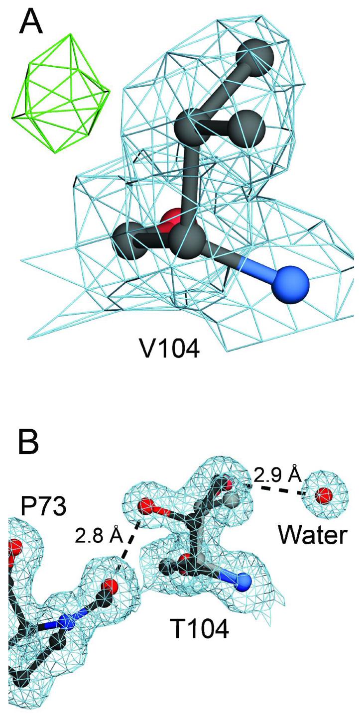
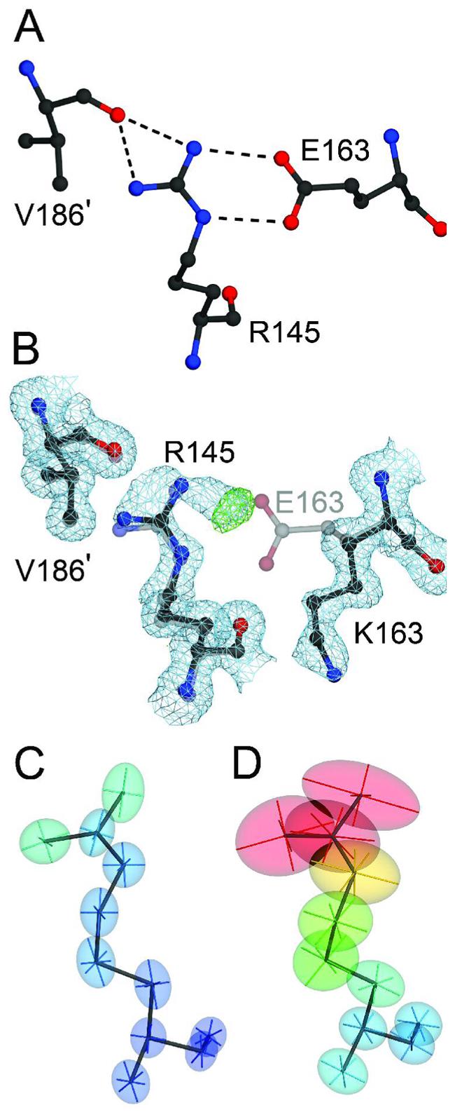
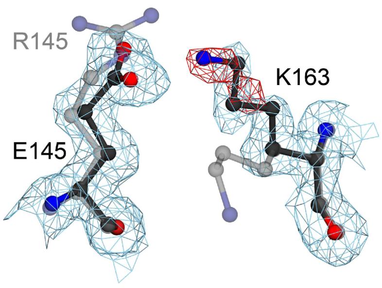
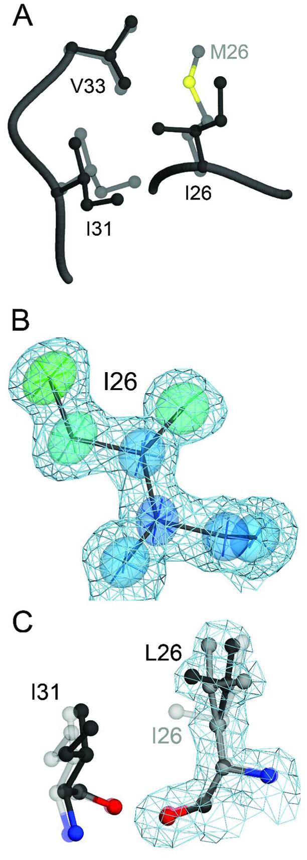
A number of missense mutations in the oxidative stress response protein DJ-1 are implicated in rare forms of familial Parkinsonism. The best-characterized Parkinsonian DJ-1 missense mutation, L166P, disrupts homodimerization and results in a poorly folded protein. The molecular basis by which the other Parkinsonism-associated mutations disrupt the function of DJ-1, however, is incompletely understood. In this study we show that three different Parkinsonism-associated DJ-1 missense mutations (A104T, E163K, and M26I) reduce the thermal stability of DJ-1 in solution by subtly perturbing the structure of DJ-1 without causing major folding defects or loss of dimerization. Atomic resolution X-ray crystallography shows that the A104T substitution introduces water and a discretely disordered residue into the core of the protein, E163K disrupts a key salt bridge with R145, and M26I causes packing defects in the core of the dimer. The deleterious effect of each Parkinsonism-associated mutation on DJ-1 is dissected by analysis of engineered substitutions (M26L, A104V, and E163K/R145E) that partially alleviate each of the defects introduced by the A104T, E163K and M26I mutations. In total, our results suggest that the protective function of DJ-1 can be compromised by diverse perturbations in its structural integrity, particularly near the junctions of secondary structural elements.


Figures

Figure 1
Figure 1
Location of three PD-associated mutations in the DJ-1 dimer. The DJ-1 dimer is represented with one monomer colored blue and the other gold. Three residues that are mutated in certain forms of familial Parkinsonism studied in this work are shown in both monomers of the DJ-1 dimer, with prime symbols indicating the symmetry-related residues. Both M26 and A104 are located in the core of dimeric DJ-1, while E163 is a surface-exposed residue in α-helix G. The figure was created with POVscript+(65).
Figure 2
Figure 2
All three PD-associated mutations thermally destabilize DJ-1. Differential scanning calorimetry (DSC) of wtDJ-1 (filled circles), M26I (open squares), A104T (filled squares) and E163K (grey triangles) shows that each pathogenic mutation reduces the apparent melting temperature (Tm) of DJ-1. The molar heat capacity at constant pressure (Cp) is calculated from the observed endotherms (ordinate), but cannot be subjected to thermodynamic analysis due to the irreversibility of DJ-1 denaturation. The Tm for each sample reported in the text is taken from the fit of a non-two state unfolding transition to the measured endotherm.
Figure 3
Figure 3
Sedimentation equilibrium ultracentrifugation of DJ-1 and PD-associated mutants. Each panel represents the measured absorbance at 277 nm as a function of radius (lower plots), and the residuals after fitting to a dimeric model for each sample (upper plots) after sedimentation equilibrium ultracentrifugation. The best-fit curve is shown as a solid line in the lower plots for each sample. Shown in panel A is wtDJ-1, panel B is A104T, panel C is E163K and panel D is M26I. Representative data for an experiment conducted at 2×10⁴ rpm and 25°C is shown. Other self-association models resulted in no improvement in the residuals and thus the simple dimer model was retained.
Figure 4
Figure 4
The secondary structural content of DJ-1 in solution is unaffected by three PD-associated mutations. Circular dichroism (CD) spectra were collected at 20°C, pH=7.5 for 10μM wtDJ-1 (filled circles), M26I (open squares), A104T (filled squares) and E163K (grey triangles) and show no significant differences in the mean residue molar ellipicity ([Θ]; ordinate) as a function of wavelength.
Figure 5
Figure 5
Buried solvent and sidechain rotameric disorder in A104T DJ-1. Panel A shows a superposition of wild-type (1SOA (5); semi-transparent grey) and A104T DJ-1 (dark grey) in the region around the site of the substitution. The introduction of a bulkier and disordered sidechain at residue 104 results in structural perturbations of the surrounding residues L72 and L112. Panel B shows a 1.05 Å resolution view of the discrete rotameric disorder at T104 and the partially occupied water molecule. Inspection of 2mFO-DFC electron density contoured at 1σ (blue) shows that the buried water molecule forms three hydrogen bonds (dashed lines) with surrounding residues, including the Oγ atom of one conformation of T104. Negative difference (mFO-DFC) electron density contoured at −3σ (red) indicates that the buried water molecule is partially occupied. This negative difference electron density disappeared when the occupancy of the water was reduced to 0.5 in the final model. The figure was created with POVscript+(65).
Figure 6
Figure 6
Hydrogen bonding stabilizes discrete disorder at T104 in A104T DJ-1. Panel A shows the environment of an engineered A104V substitution in DJ-1. Inspection of 1.85 Å resolution 2mFo-DFc electron density at 1.0σ (blue) shows that V104 is less disordered than T104, despite the steric similarity of these two residues. Positive difference (mFo-DFc) electron density contoured at 3.0σ (green), however, indicates that the residue likely samples a second minor conformation. In panel B, T104 makes two mutually exclusive hydrogen bonds that favor discrete disorder for this residue. The higher occupancy conformation for T104 is shown in the darker line, electron density (2mFo-DFc) is contoured at 1.0σ (blue), and hydrogen bonds are shown in dashed lines with their distances provided in Ångstroms. The orientation of residue 104 in panels A and B is the same. The figure was created with POVscript+(65).
Figure 7
Figure 7
The E163K substitution disrupts a critical salt bridge in DJ-1. Panel A shows the network of hydrogen bonds (dashed lines) that extend from the E163-R145 salt bridge to V186 in the other monomer of the DJ-1 dimer (marked with a prime). Panel B shows a superposition of wild-type (1SOA(5); semi-transparent grey) and the 1.15 Å resolution crystal structure of E163K DJ-1 (dark grey), showing that the substitution of a lysine at position 163 disrupts the salt bridge with R145 and leads to reorientation of the K163 sidechain. Electron density (2mFO-DFC) contoured at 1σ (blue) and positive difference mFO-DFC electron density contoured at 4σ (green) calculated from the E163K DJ-1 model indicate that loss of the E163-R145 salt bridge leads to disorder at Arg145 in E163K DJ-1. The increased mobility of R145 is evident by comparison of the thermal ellipsoids calculated from the refined ADPs of wtDJ-1 (Panel C) and E163K DJ-1 (Panel D). The thermal ellipsoids with their principal axes are shown at the 50% probability level and are colored to indicate the magnitude of the total atomic displacement (blue; 6 Å2, red; 40 Å2). The figure was created with POVscript+(65).
Figure 8
Figure 8
An engineered E163K/R145E double mutation creates a poorly ordered salt bridge. A superposition of E163K (semi-transparent grey) and an engineered E163K/R145E DJ-1 double mutant (darker line) shows that restoring electrostatic complementarity creates a salt bridge with the poorly ordered sidechain of K163. Electron density (2mFO-DFC) calculated from the 1.5 Å resolution crystal structure of E163K/R145E DJ-1 is contoured at 1σ (blue) and shows that K163 moves to interact with E145. Prominent negative difference (mFO-DFC) electron density contoured at −3.5σ (red) indicates that K163 is poorly ordered and that native hydrogen bonding interactions between R145 and E163 in wtDJ-1 are required for optimal interactions in this region of the protein. The figure was created with POVscript+(65).
Figure 9
Figure 9
The M26I substitution results in packing defects in the core of DJ-1. Panel A shows a superposition of wild-type (1SOA(5); semi-transparent grey) and M26I DJ-1 (dark grey) in the region around the site of the substitution. The M26I substitution results in a ∼0.7 Å displacement of I31 to accommodate the Cγ2 atom of I26. In addition, the loss of the Sδ and Cε atoms of M26 creates a minor packing defect in the hydrophobic core of the protein. In Panel B, 2mFO-DFC electron density contoured at 1σ (blue) is shown with thermal ellipsoids calculated from the refined ADPs of I26, indicating that this residue is well-ordered in the crystal. The thermal ellipsoids are shown at the 60% probability level with their principal axes and are colored to indicate the magnitude of the total atomic displacement (blue; 6 Å2, red; 15 Å2). Panel C shows a superposition of M26I (semi-transparent grey) and the engineered M26L substitution, with alternate conformations for L26 shown in black and dark grey. The cavity created by the M26L substitution allows L26 to sample two rotameric conformations in the core of DJ-1, as shown by 2mFO-DFC electron density contoured at 1σ (blue) calculated from the 1.5 Å resolution crystal structure of M26L DJ-1. The figure was created with POVscript+((65).

References

    1. Bonifati V, Rizzu P, van Baren MJ, Schaap O, Breedveld GJ, Krieger E, Dekker MC, Squitieri F, Ibanez P, Joosse M, van Dongen JW, Vanacore N, van Swieten JC, Brice A, Meco G, van Duijn CM, Oostra BA, Heutink P. Mutations in the DJ-1 gene associated with autosomal recessive early-onset parkinsonism. Science. 2003;299:256–259. - PubMed
    1. Nagakubo D, Taira T, Kitaura H, Ikeda M, Tamai K, Iguchi-Ariga SM, Ariga H. DJ-1, a novel oncogene which transforms mouse NIH3T3 cells in cooperation with ras. Biochem. Biophys. Res. Commun. 1997;231:509–513. - PubMed
    1. Bandyopadhyay S, Cookson MR. Evolutionary and functional relationships within the DJ1 superfamily. BMC Evol. Biol. 2004;4 - PMC - PubMed
    1. Lucas JI, Marin I. A New Evolutionary Paradigm for the Parkinson Disease Gene DJ-1. Mol. Biol. Evol. 2006;24:551–561. - PubMed
    1. Canet-Aviles RM, Wilson MA, Miller DW, Ahmad R, McLendon C, Bandyopadhyay S, Baptista MJ, Ringe D, Petsko GA, Cookson MR. The Parkinson's disease protein DJ-1 is neuroprotective due to cysteine-sulfinic acid-driven mitochondrial localization. Proc. Natl. Acad. Sci. USA. 2004;101:9103–9108. - PMC - PubMed

Publication types

MeSH terms

Substances