Review
. 2008 Apr 1;111(7):3331-42.
doi: 10.1182/blood-2007-10-052761. Epub 2008 Jan 8.

Zebrafish in hematology: sushi or science?

Affiliations
Review

Zebrafish in hematology: sushi or science?

Duncan Carradice et al. Blood. .

Abstract

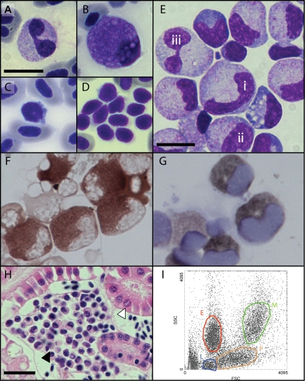
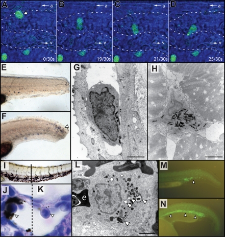
After a decade of the "modern era" of zebrafish hematology research, what have been their major contributions to hematology and what challenges does the model face? This review argues that, in hematology, zebrafish have demonstrated their suitability, are proving their utility, have supplied timely and novel discoveries, and are poised for further significant contributions. It presents an overview of the anatomy, physiology, and genetics of zebrafish hematopoiesis underpinning their use in hematology research. Whereas reverse genetic techniques enable functional studies of particular genes of interest, forward genetics remains zebrafish's particular strength. Mutants with diverse and interesting hematopoietic defects are emerging from multiple genetic screens. Some mutants model hereditary blood diseases, occasionally leading to disease genes first; others provide insights into developmental hematology. Models of malignant hematologic disorders provide tools for drug-target and pharmaceutics discovery. Numerous transgenic zebrafish with fluorescently marked blood cells enable live-cell imaging of inflammatory responses and host-pathogen interactions previously inaccessible to direct observation in vivo, revealing unexpected aspects of leukocyte behavior. Zebrafish disease models almost uniquely provide a basis for efficient whole animal chemical library screens for new therapeutics. Despite some limitations and challenges, their successes and discovery potential mean that zebrafish are here to stay in hematology research.


Figures

Figure 1
Figure 1
Hematopoietic specification and specific cell lineages in the zebrafish embryo. (A-E) Markers of hematopoietic specification. (A) The earliest site of primitive hematopoiesis can be identified in the posterior lateral plate mesoderm using a riboprobe for tal1(scl) (blue, open arrowhead). (B) gata1 expression (blue) in the developing intermediate cell mass (ICM). (C) spi1 (pu.1) expression (blue) in the anterior lateral plate mesoderm (open arrowhead), below the head (closed arrowhead), defines the region producing the first wave of primitive macrophages. (D,E) Definitive hematopoiesis, marked by cmyb and runx1 (blue), commences in the ventral wall of the dorsal aorta (arrowheads). (F-K) Markers of specific hematopoietic cell lineages. (F) Lineage-committed erythroid cells expressing hbbe3 (globinβe3) (blue) in the ICM and circulating cells over the yolk. (G) mpx-expressing granulocytes (blue) in circulation over the yolk and in the ventral tail (open arrowheads). (H) Sudan black cytochemistry marks the same dispersed granulocyte population (open arrowheads). (I) ighm (IgM) expression in a B lymphocyte (open arrowhead) in a kidney section, adjacent to a renal tubule (closed arrowhead). (J) T lymphocytes marked by rag1 expression (blue) in the bilateral thymi. (K) CD41 (itga2b) expression (blue) in circulating thrombocytes over the yolk (open arrowhead). (A-G,I-K) Gene expression by whole mount in situ hybridization. Microscopy was performed using a Nikon SMZ-1500 microscope (Nikon, Melville, NY) equipped with a 0.75-11.25× objective (A-H,J-K) and a Nikon Optiphot-2 microscope equipped with a 100×/1.40 oil objective (I). Images were obtained using a Zeiss Axiocam MRc5 digital camera (Carl Zeiss, Thornwood, NY) with Axiovision AC software (Release 4.5). Images were processed using Adobe Photoshop CS2 9.0 and Adobe Illustrator CS2 12.0.1 (Adobe Systems, San Jose, CA).
Figure 2
Figure 2
Morphology of adult zebrafish hematopoietic cells. (A-D) Peripheral blood smears showing: (A) bilobed neutrophil, (B) eosinophil, (C) lymphocyte and nucleated erythrocytes, and (D) aggregate of thrombocytes with visible cytoplasmic projections. (E-G) Kidney marrow cell cytospin showing: (E) progression of granulocyte maturation from immature (i,ii) to mature (iii) forms, (F) identification of granulocytes by myeloperoxidase cytochemistry, and (G) identification of granulocytes by Sudan black cytochemistry. (H) A cluster of hematopoietic cells (closed arrowhead) nestled between renal tubules (open arrowhead) (hematoxylin and eosin stained section). (I) FACS analysis of kidney marrow cells by forward scatter (FSC) and side scatter (SSC) separates erythroid (red), myelomonocytic (green), lymphocytic (blue), and progenitor (orange) cell populations. Bars represent 10μm (A-G) and 20μm (H). Microscopy was performed using a Nikon Optiphot-2 microscope equipped with a 40×/1.0 and 100×/1.40 oil objective. Images were obtained using a Zeiss Axiocam MRc5 digital camera (Carl Zeiss) with Axiovision AC software (Release 4.5). Images were processed using Adobe Photoshop CS2 9.0 and Adobe Illustrator CS2 12.0.1 (Adobe Systems).
Figure 3
Figure 3
Demonstrations of hematologic cell function in zebrafish embryos. (A-D) Using spi1:EGFP transgenic animals, in vivo time-lapse confocal microscopy over 30 seconds shows: (A) migration of a leukocyte (arrowhead) from the arteriole (a) and its movement between cells of the extravascular compartment toward a venule (v) (B-D). Video S1 is the movie from which these 4 still images were selected; it displays more clearly the leukocyte assuming a “dumbbell” morphology as it passes between extravascular supporting cells. (E,F) Neutrophil chemotaxis mpx-expressing neutrophils (by whole mount in situ hybridization, blue) are not abundant at the site of injury 1 hour after tail transection (E) but accumulate at the injured site after 8 hours (F, arrowhead). (G,H) By electron microscopy of 7 dpf embryos, neutrophils identified by their pathognomonic electron-dense granules are demonstrated within the vasculature (G) and muscle (H) in the vicinity of a sterile wound. (I-L) Macrophage phagocytic function. After injection of India ink, there is nonembolic accumulation of carbon particles in the ventral tail (I), within a marginated phagocytic cell (open arrowhead, J), in a field including a marginated bilobed granulocyte (K) for comparison. Dashed line divides different focal planes of the same tissue section. Electron microscopy demonstrates phagosomes (open arrowheads) within an adult splenic macrophage (L). “e” indicates an adjacent erythrocyte; I is a brightfield unstained image; J and K are hematoxylin and eosin-stained sections. (M,N) Expansion of hematopoiesis by jak2a overexpression. Normal fluorescence of the ICM (arrowhead) in Tg(spi1:EGFP) embryo at 24 hpf (M). Expansion of the ICM (arrowheads) is demonstrated after injection of constitutively active zebrafish tel-jak2a (N). Bars represent 2 μm (G,H,L). Microscopy was performed using a Bio-Rad MRC1024 confocal microscope (Bio-Rad, Hercules, CA; A-D), Siemens Elmiscope 102 transmission electron microscope (Siemens, Munich, Germany; G,H,L), Nikon SMZ-1500 microscope (Nikon) equipped with a 0.75-11.25× objective (E,F,I), a Nikon Optiphot-2 microscope equipped with a 100×/1.40 oil objective (J,K) and a Leica MZFIII fluorescence microscope (Leica, Wetzlar, Germany) equipped with a 0.8-10.0× objective (M,N). Electron microscopy images were printed to photographic paper and then digitised. Images were obtained using a Zeiss Axiocam MRc5 digital camera (Carl Zeiss) with Axiovision AC software (Release 4.5) or an Olympus DP70 digital camera (Olympus-Australia, Melbourne, Australia) with DP controller 1.2.1.108 software. Images were processed using Adobe Photoshop CS2 9.0 and Adobe Illustrator CS2 12.0.1 (Adobe Systems).
Figure 4
Figure 4
Zebrafish hematopoietic mutants. (A) cloche, a mutant in the earliest acting gene in hematopoiesis, upstream of the hemangioblast marker tal1(scl), shows a lack of circulating red hemoglobinized erythrocytes circulating over the yolk compared with wild-type (arrowheads) in brightfield images (left panels), and lacks expression of gata1 (red, erythrocytes) and lcp(l-plastin) (blue, myelomonocytes) in 2-color whole mount in situ hybridization (right panels). (B) The dorsalized alk8 mutant laf gl2 lacks anterior expression of the early myeloid marker spi1(pu.1). (C) The ventralized chordin mutant dino has expansion of the intermediate cell mass with increased expression of gata1 (blue). (D) Blood smears comparing wild-type and retsina (a scl4a1 (erythrocyte band 3) mutant) erythrocytes; retsina erythrocytes have distinctive binucleate erythroblasts (closed arrowhead), reminiscent of congenital dyserythropoietic anemia type II. (A-C) Gene expression by whole mount in situ hybridization. Microscopy was performed using a Nikon SMZ-1500 microscope (Nikon) equipped with a 0.75-11.25× objective (A-C). Images were obtained using a Zeiss Axiocam MRc5 digital camera (Carl Zeiss), with Axiovision AC software (Release 4.5). Images were processed using Adobe Photoshop CS2 9.0 and Adobe Illustrator CS2 12.0.1 (Adobe Systems).

Similar articles

Cited by

References

    1. Roosen-Runge E. Observations of the early development of the zebra fish, Brachydanio rerio. Anat Rec. 1937;70(Suppl 1):103.
    1. Laale HW. The biology and use of zebrafish, Brachydanio rerio, in fisheries research: a literature review. J Fish Biol. 1977;10:121–173.
    1. Colle-Vandevelde A. Blood anlage in Teleosti. Nature. 1963;198:1223. - PubMed
    1. Rieb J-P. La Circulation Sanguine Chez L'Embryon De Brachydanio Rerio. Ann Embryol Morphogenese. 1973;6:43–54.
    1. Johnson RA, Hart NH, Powell D. Studies on the morphology of blood cells in the fresh water teleost, Brachydanio rerio. Bull N Y Acad Sci. 1973;18:20–21.

Publication types

MeSH terms