. 2008 Apr 15;111(8):4386-91.
doi: 10.1182/blood-2007-10-115725. Epub 2008 Jan 8.

Donor cell-derived osteopoiesis originates from a self-renewing stem cell with a limited regenerative contribution after transplantation

Affiliations

Donor cell-derived osteopoiesis originates from a self-renewing stem cell with a limited regenerative contribution after transplantation

Massimo Dominici et al. Blood. .

Abstract

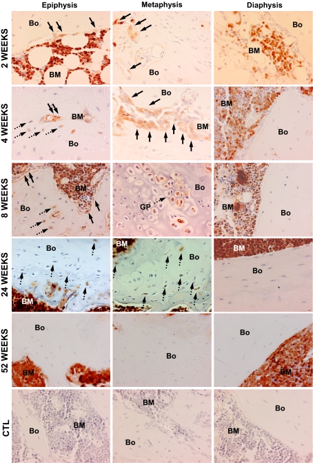
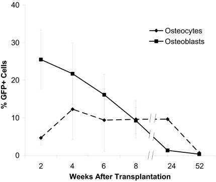
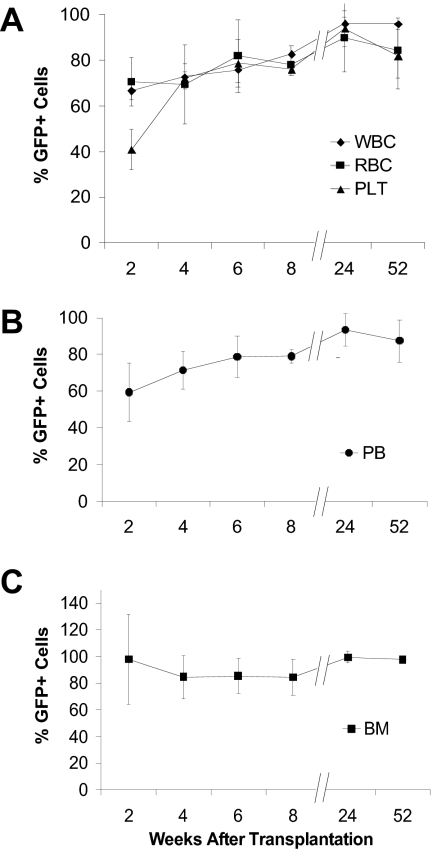
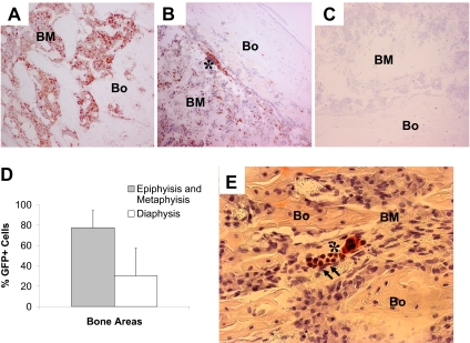
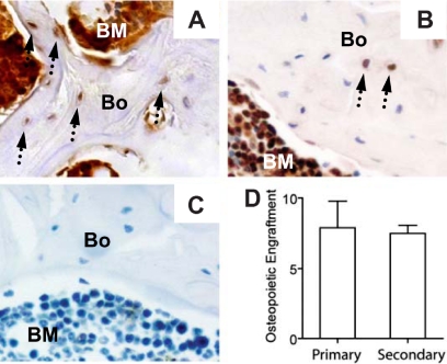
In principle, bone marrow transplantation should offer effective treatment for disorders originating from defects in mesenchymal stem cells. Results with the bone disease osteogenesis imperfecta support this hypothesis, although the rate of clinical improvement seen early after transplantation does not persist long term, raising questions as to the regenerative capacity of the donor-derived mesenchymal progenitors. We therefore studied the kinetics and histologic/anatomic pattern of osteopoietic engraftment after transplantation of GFP-expressing nonadherent marrow cells in mice. Serial tracking of donor-derived GFP(+) cells over 52 weeks showed abundant clusters of donor-derived osteoblasts/osteocytes in the epiphysis and metaphysis but not the diaphysis, a distribution that paralleled the sites of initial hematopoietic engraftment. Osteopoietic chimerism decreased from approximately 30% to 10% by 24 weeks after transplantation, declining to negligible levels thereafter. Secondary transplantation studies provided evidence for a self-renewing osteopoietic stem cell in the marrow graft. We conclude that a transplantable, primitive, self-renewing osteopoietic cell within the nonadherent marrow cell population engrafts in an endosteal niche, like hematopoietic stem cells, and regenerates a significant fraction of all bone cells. The lack of durable donor-derived osteopoiesis may reflect an intrinsic genetic program or exogenous environmental signaling that suppresses the differentiation capacity of the donor stem cells.


Figures

Figure 1
Figure 1
Nonadherent donor bone marrow cells engraft as osteoblasts and osteocytes after transplantation. (A) Representative photomicrograph of a bone/bone marrow section taken from a mouse after nonadherent marrow cell transplantation double stained with anti-GFP (black) and antiosteocalcin (red) antibodies. Several double-positive donor osteoblasts are distributed along the surface of endosteal bone, with a donor (GFP+) osteocyte embedded in bone. Osteocalcin-expressing host cells (H; red stain without GFP) are indicated by arrows. (B) Bone/bone marrow section double stained with anti-GFP (black) and anti-collagen l (red) antibodies. Donor osteoblasts and osteocytes were detected by red/black colocalization. Collagen-expressing host osteocytes (H) are indicated by arrows. (C) Control bone section from a mouse transplanted with nontransduced nonadherent bone marrow cells and stained with anti-GFP and isotype control antibodies. Original magnification for all panels, 1600× (400× optical, 4× digital).
Figure 2
Figure 2
Patterns of bone engraftment with increasing time after transplantation of nonadherent bone marrow cells. Sections taken at 2 to 52 weeks after transplantation from different regions of bone (Bo) and bone marrow (BM) were stained with anti-GFP antibody (red). At 2 weeks, GFP+ osteoblasts (arrows) engrafted as clusters next to host (GFP) osteoblasts in the epiphysis and in the metaphysis, while in the diaphysis the osteoblasts remained almost exclusively host derived. Beginning at 4 weeks, GFP+ osteocytes (dashed arrows) were consistently detected in both the metaphysis and epiphysis. At 8 weeks, in the metaphysis, in particular, Bo-embedded GFP+ cells could be identified under the growth plate (GP) as cell clusters. Again, the donor contribution was virtually undetectable in the diaphysis. At 24 weeks, a similar pattern of donor-derived osteopoiesis was evident. By 52 weeks, neither donor Bo nor BM contained GFP+ osteoblasts or osteocytes. The bottom row consists of photomicrographs of control (CTL) Bo/BM sections from a mouse that received a transplant of untransduced cells that were stained with anti-GFP antibody. Original magnification of all panels, 400×.
Figure 3
Figure 3
Kinetics of bone engraftment after transplantation. Robust GFP+ osteoblast engraftment was detected at 2 and 4 weeks after transplantation with significant declines thereafter: 6 weeks (P = .02), 8 weeks (P = .04), 24 weeks (P = .01). GFP+ osteocyte engraftment showed a different kinetic profile characterized by a significant increase at 4 weeks (P = .04), a plateau phase and a significant decrease to a negligible level at 52 weeks (P = .004). To quantify the engraftment of donor GFP + cells in bone, we scored 20 random 400× fields in both the epiphysis and the metaphysis of 10 bone sections taken from each mouse at different times after transplantation (n ≥ 4 mice per group). Experiments were performed in triplicate. The reported values are mean (+SD) percentages of GFP+ cells per 400× microscopic field.
Figure 4
Figure 4
Nonadherent bone marrow cells can produce stable hematopoietic engraftment. (A) GFP expression by white blood cells (WBC), red blood cells (RBC), and platelets (PLT) assessed by flow cytometry with the whole peripheral blood analyses. The findings are reported as mean (+SD) percentages of GFP+ cells representing each of the 3 hematopoietic lineages. Values for all lineages were significantly increased at 24 weeks (WBC, P < .001; RBC, P = .03; PLT, P = .008) compared with 2 weeks, becoming stable thereafter. (B) Flow cytometric analysis of GFP expression in whole peripheral blood (PB) collected from the mice that underwent transplantation (n ≥ 4 per group) at increasing times after transplantation. The findings are reported as mean (+SD) percentages of GFP+ cells. (C) GFP expression in bone marrow assessed at different times after transplantation. We scored 20 randomly selected 400× fields in representative sections from each mouse (n ≥ 4 mice per group). The values were derived from immunohistochemical analyses of bone/bone marrow sections stained with anti-GFP antibody and are expressed as mean (+SD) percentages of GFP+ cells in each microscopic field. All experiments were performed in triplicate.
Figure 5
Figure 5
Nonadherent bone marrow cells engraft preferentially in metaphysis and epiphysis in the early regenerative phases after BMT. (A) Photomicrograph of a bone (Bo)/bone marrow (BM) section taken from the epiphysis of a mouse that underwent transplantation killed at 2 weeks after transplantation and stained with anti-GFP antibody (red). Original magnification, 100×. (B) Low level of GFP+ cells engrafted as clusters in the diaphysis, mainly in close proximity to the endosteal bone surface (asterisk). (C) Section of bone from a mouse that received a transplant of untransduced nonadherent marrow cells and stained with anti-GFP antibody (negative control). (D) Comparison of GFP+ engraftment in the diaphysis versus the metaphysis/epiphysis at 2 weeks after transplantation. A total of 20 random 400× fields of bone sections from each of 4 mice were studied. The reported values are mean (+SD) percentages of GFP+ cells per field. The difference in engraftment is highly significant (P < .001). (E) Photomicrograph of a bone (Bo)/bone marrow (BM) section taken from the metaphysis of a transplanted mouse killed at 2 days after transplantation and stained with anti-GFP antibody (red). The cluster of early osteopoietic engraftment (arrows) is adjacent to donor (GFP+) hematopoietic cells (asterisk). Original magnification, 400×.
Figure 6
Figure 6
The osteopoietic capacity of nonadherent bone marrow cells is preserved in secondary recipients. Immunohistochemical identification of donor-derived bone cells (arrows) in primary (A) and secondary (B) recipients. (C) Section of bone from a mouse that received a transplant of untransduced nonadherent marrow cells and stained with anti-GFP antibody (negative control). (D) Comparison of the percentages of donor-derived osteopoiesis in primary (n = 3 mice) versus secondary (n = 10 mice) recipients. The reported values are mean (± SD) percentages of GFP+ cells. Bone engraftment was quantified as described in Figure 3; P > .05.

Comment in

Similar articles

Cited by

References

    1. Nilsson SK, Dooner MS, Weier HU, et al. Cells capable of bone production engraft from whole bone marrow transplants in nonablated mice. J Exp Med. 1999;189:729–734. - PMC - PubMed
    1. Horwitz EM, Prockop DJ, Fitzpatrick LA, et al. Transplantability and therapeutic effects of bone marrow-derived mesenchymal cells in children with osteogenesis imperfecta. Nat Med. 1999;5:309–313. - PubMed
    1. Horwitz EM, Gordon PL, Koo WKK, et al. Isolated allogeneic bone marrow-derived mesenchymal cells engraft and stimulate growth in children with osteogenesis imperfecta: implications for cell therapy of bone. Proc Natl Acad Sci USA. 2002;99:8932–8937. - PMC - PubMed
    1. Brazelton TR, Nystrom M, Blau HM. Significant differences among skeletal muscles in the incorporation of bone marrow-derived cells. Dev Biol. 2003;262:64–74. - PubMed
    1. Dominici M, Pritchard C, Garlits JE, Hofmann T, Persons DA, Horwitz EM. Hematopoietic cells and osteoblasts are derived from a common marrow progenitor after bone marrow transplantation. Proc Natl Acad Sci U S A. 2004;101:11761–11766. - PMC - PubMed

Publication types