. 2008 Jan 16;100(2):140-54.
doi: 10.1093/jnci/djm288. Epub 2008 Jan 8.

Kidney-targeted Birt-Hogg-Dube gene inactivation in a mouse model: Erk1/2 and Akt-mTOR activation, cell hyperproliferation, and polycystic kidneys

Affiliations

Kidney-targeted Birt-Hogg-Dube gene inactivation in a mouse model: Erk1/2 and Akt-mTOR activation, cell hyperproliferation, and polycystic kidneys

Masaya Baba et al. J Natl Cancer Inst. .

Abstract

Background: Patients with Birt-Hogg-Dubé (BHD) syndrome harbor germline mutations in the BHD tumor suppressor gene that are associated with an increased risk for kidney cancer. BHD encodes folliculin, a protein that may interact with the energy- and nutrient-sensing 5'-AMP-activated protein kinase-mammalian target of rapamycin (AMPK-mTOR) signaling pathways.

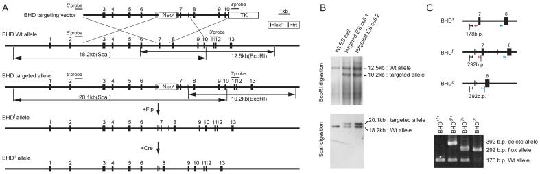
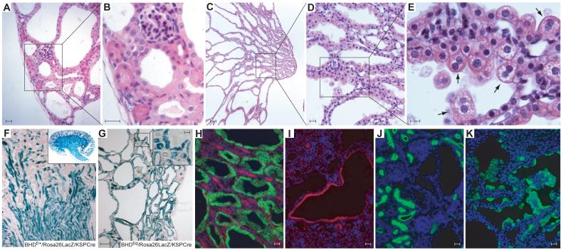
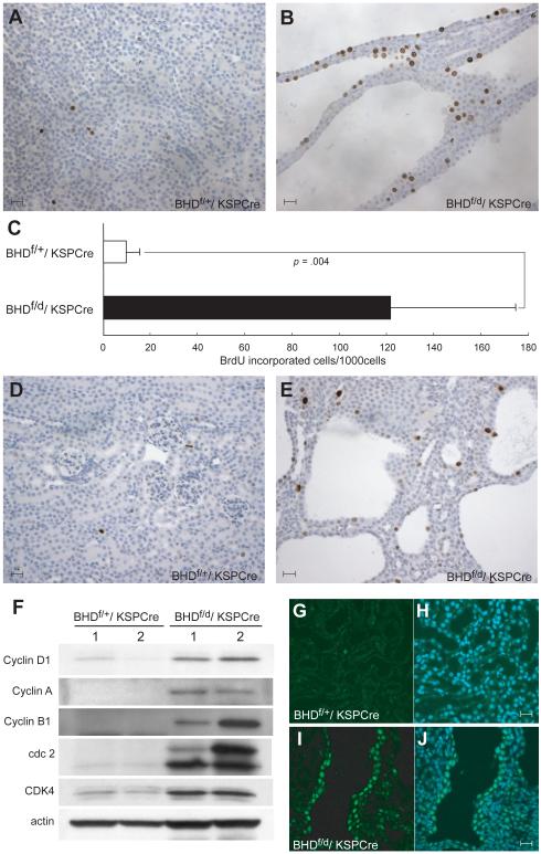
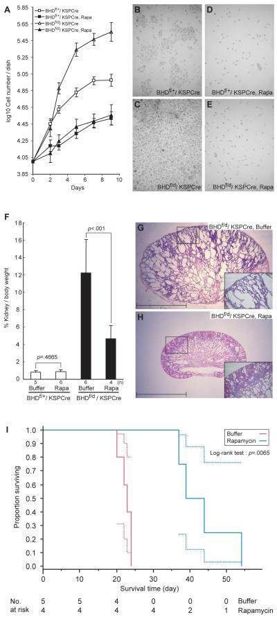
Methods: We used recombineering methods to generate mice with a conditional BHD allele and introduced the cadherin 16 (KSP)-Cre transgene to target BHD inactivation to the kidney. Kidney cell proliferation was measured by BrdU incorporation and phospho-histone H3 staining. Kidney weight data were analyzed with Wilcoxon's rank-sum, Student's t, and Welch's t tests. Hematoxylin and eosin staining and immunoblot analysis and immunohistochemistry of cell cycle and signaling proteins were performed on mouse kidney cells and tissues. BHD knockout mice and kidney cells isolated from BHD knockout and control mice were treated with the mTOR inhibitor rapamycin. Mouse survival was evaluated by Kaplan-Meier analyses. All statistical tests were two-sided.

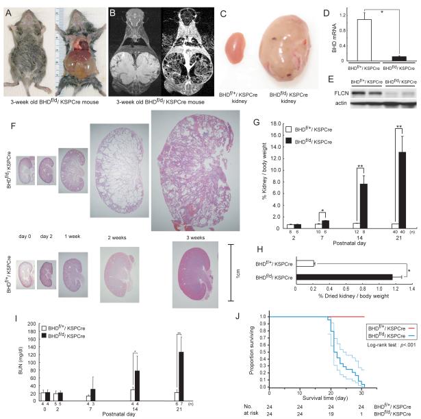
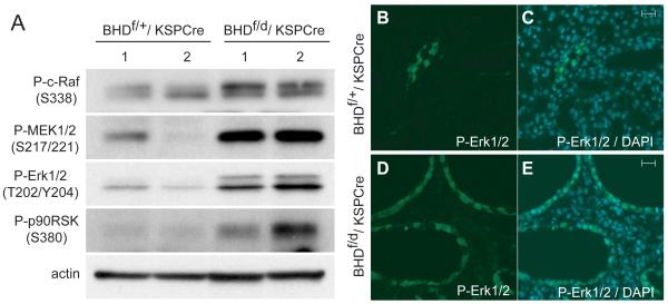
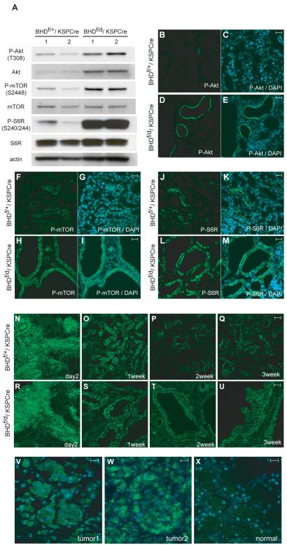
Results: BHD knockout mice developed enlarged polycystic kidneys and died from renal failure by 3 weeks of age. Targeted BHD knockout led to the activation of Raf-extracellular signal-regulated protein kinase (Erk)1/2 and Akt-mTOR pathways in the kidneys and increased expression of cell cycle proteins and cell proliferation. Rapamycin-treated BHD knockout mice had smaller kidneys than buffer-treated BHD knockout mice had (n = 4-6 mice per group, relative kidney/body weight ratios, mean = 4.64% vs 12.2%, difference = 7.6%, 95% confidence interval = 5.2% to 10.0%; P < .001) and longer median survival time (n = 4-5 mice per group, 41.5 vs 23 days; P = .0065 ).

Conclusions: Homozygous loss of BHD may initiate renal tumorigenesis in the mouse. The conditional BHD knockout mouse may be a useful research model for dissecting multistep kidney carcinogenesis, and rapamycin may be considered as a potential treatment for Birt-Hogg-Dubé syndrome.


Figures

Fig. 1
Fig. 1. Generation of conditional BHD knockout mice
A) Targeting strategy. Birt-Hogg-Dube’gene (BHD) targeting vector was constructed by recombineering methodology using homologous recombination (27). A neomycin resistance (Neor) cassette flanked by Frt (bar) and loxP (triangle) sequences was inserted into intron 6 for positive selection, and the thymidine kinase gene was included for negative selection. A second loxP sequence was inserted into intron 7. Correctly-targeted embryonic stem (ES) cells were identified by Southern blot analysis and injected into blastocysts to produce chimeras. Backcrossing to C57BL/6 mice produced heterozygous F1 offspring with germline transmission of the BHD floxed (f)-Neo allele. The Neo cassette flanked by Frt sites was excised in vivo by crossing with mice expressing the Flp recombinase transgene under the ubiquitous β-actin promoter. To produce the BHD deleted (d) allele, BHD f/+ mice were crossed with mice expressing the Cre recombinase transgene under the ubiquitous β-actin promoter (24). Deletion of exon 7 resulted in a frame shift and premature termination codon in exon 8, which caused mRNA degradation by the nonsense-mediated decay mRNA surveillance system (43). B) The targeted ES cells were screened by Southern blotting of EcoRI and ScaI digested DNA using two different external probes located outside the targeting sequence as shown in (A). C) Polymerase chain reaction (PCR)-based genotyping was performed using DNA extracted from mouse tails for routine monitoring of inheritance in offspring. Locations of PCR primers are indicated by arrows.
Fig. 2
Fig. 2. Phenotypic features of BHD-targeted deletion in the kidney
A) Gross picture of a 3-week old BHDf/d/KSP (cadherin 16 kidney-specific promoter)-Cre mouse shows a distended abdomen (left panel). The large cystic kidneys fill the abdominal cavity (right panel) and are found in 100% of BHDf/d/KSP-Cre mice. B) T2 weighted coronal magnetic resonance imaging (MRI) image (left) and corresponding Gadolinium-enhanced T1 weighted dynamic subtraction MRI image (right) of a 3-week-old BHDf/d/KSP-Cre mouse show enlarged cystic kidneys with reticular interstitium and delayed excretion of contrast medium. C) Comparison of gross features of 3-week-old control BHDf/+/KSPCre and knockout BHDf/d/ KSP-Cre kidneys reveals a normal phenotype in control mice. Knockout BHDf/d/ KSP-Cre kidneys show symmetric enlargement without focal masses. One representative image of 40 mice for each genotype is shown. D) BHD mRNA expression in kidneys of 3-week-old mice was quantified by quantitative reverse transcription-polymerase chain reaction (qRT-PCR) using exon 6 and 7 amplification. Three mice of each genotype were analyzed. Data are presented as means and 95% confidence intervals from three independent experiments performed in triplicate (Welch’s t test:*P<.001). E) Folliculin (FLCN) expression in control and knockout kidneys was estimated by immunoblotting. One representative of three independent experiments performed in two mice is shown. F) Comparison of BHD control and knockout kidney histology (hematoxylin and eosin staining) at different ages shows no obvious differences at days 0 and 2. At 1 week, BHD-knockout mice have enlarged kidneys with dilated collecting ducts and a few dilated cortical tubules. By 2 weeks of age, most collecting ducts in the medulla are markedly dilated. The entire BHD-knockout kidney is diffusely filled with cystic collecting ducts and tubules at 3 weeks of age, and at this point anatomic distinction between cortex and medulla is lost. One representative of at least three mice at each age is shown. G) Relative ratio of kidney to body weight (BW;100 X kidney weight/[BW—kidney weight]) was calculated at different ages. The mean kidney weight of the BHDf/d/KSP-Cre mice at 3 weeks of age was 13.1% of body weight compared with 0.77% for control mice (n=40 for each group, difference=12.3%, 95% CI=11.1% to 13.6%; Welch’s t test:P<.001). Data are represented as means and 95% confidence intervals (*P =.002, ** P <.001). H) Relative ratio of dried kidney to body weight (100 X dried kidney weight/[BW—wet kidney weight]) of 3-week-old mice was calculated. Even with loss of cystic fluid, the dry weight of BHDf/d/KSP-Cre kidneys was statistically significantly greater than control kidneys (mean=1.16%, n=8 versus mean=0.21%, n=10, difference=0.95%, 95% CI=0.84% to 1.06%; Welch’s t test: * P<.001). Data are represented as means and 95% confidence intervals. I) BHDf/d/KSP-Cre mice die of renal failure. Blood urea nitrogen (BUN) levels were determined at different ages. Statistically significant elevation of BUN levels was observed at 2 weeks of age in BHDf/d/KSP-Cre mice compared with littermate controls (n=4 for each group, mean=77.7 mg/dL versus mean=29.2mg/dL, difference=48.5mg/dL, 95% CI=18.6 to 78.3 , Welch’s t test:*P=.03). Similar statistically significant differences were seen between 3-week old BHDf/d/KSP-Cre mice (n=7) and control littermates (n=6) (mean=126.1 mg/dL versus mean=21.8 mg/dL, difference=104.3 mg/dL, 95% CI=64.9 to 143.9 , Welch’s t test:**P=.0012). Data are represented as means and 95% confidence intervals. J) Kaplan-Meier survival analysis shows a statistically significant difference between control and BHD knockout mice (log-rank test, P<.001). Median survival time of BHD knockout mice is 21.5 days (n=24).
Fig.3
Fig.3. Histology and immunostaining of kidneys of BHD knockout mice
A–E) Histology of hematoxylin and eosin stained kidney from a 3-week-old BHDf/d/KSP-Cre mouse. A) The subcapsular region of the kidney shows dilated distal tubules, mildly compressed glomeruli and relatively normal proximal tubules. One representative image of ten sections is shown. B) Higher magnification of (A). Bowman’s space is minimal and glomerular tufts are mildly compressed. Morphologically normal proximal tubules are surrounded by dilated distal tubules lined by hypertrophic cells with enlarged eosinophilic granular cytoplasm and enlarged nuclei. C) Collecting ducts in the medulla are severely dilated. D-E) Higher magnification of medulla. Note hypertrophic cells with enlarged eosinophilic, granular cytoplasm that protrude into the lumen and an occasional binucleated cell (arrow). F) To confirm the Cre expression pattern in the kidney, BHDf/f/Rosa26lacZ mice were generated and crossed with BHDd/+/KSP-Cre mice. X-Gal staining of a BHDf/+/Rosa26lacZ/KSP-Cre mouse kidney at 3 weeks of age shows strong staining in the medulla and scattered staining pattern in the cortex. G) All dilated tubules of 3-week-old BHDf/d/Rosa26lacZ/KSP-Cre mouse kidneys show strong X-Gal staining. Morphologically normal proximal tubules also show mosaic staining (insert). H) Dolichos biflorus agglutinin (DBA; green) and Na-K-Cl cotransporter 2 (NKCC2; red) staining in 1-week-old BHDf/d/KSP-Cre mouse kidneys. Dilated tubules are DBA (collecting duct marker) positive. Loops of Henle, which are NKCC2 positive, are morphologically normal. I) Dilated tubules in the cortex are thiazide-sensitive Na-Clcotransporter (TSC; red) positive, a marker of distal tubule. J) Lotus tetragonolobus agglutinin (LTA; green) staining of BHDf/d/KSP-Cre mouse kidneys identifies apparently normal proximal tubules but does not stain dilated tubules. K) Markedly hypertrophic cells lining the dilated ducts of 2-week-old BHD knockout mouse kidneys are stained by the intercalated cell marker, vacuolar H+-ATPase (green). Scale bar=20 μm for A,B,D,H,I,J,K; 10 μm for E; 100 μm for C,F,G.
Fig. 4
Fig. 4. Evidence of hyperproliferating cells in the kidney of BHDf/d/ KSP-Cre mice
5′-Bromo-2′-deoxyouridine (BrdU) (100 μg/g body wt) was injected intraperitoneally at P14 into BHDf/+/KSP-Cre and BHDf/d/KSP-Cre mice 2 hr before euthanization. A) BrdU staining in BHDf/+/KSP-Cre mouse kidney. B) BrdU staining in BHDf/d/KSP-Cre mouse kidney. C) Greater than 10-fold more BrdU incorporation was seen in BHDf/d/KSP-Cre mouse kidneys compared with BHDf/+/KSP-Cre mouse kidneys (n=5 for each group, mean=121.8 per 1000 cells versus 9.6 per 1000 cells, difference=112.2, 95% CI=59.3 to 165.0; Welch’s t test:P=.004). 1000 cells per field were counted in five randomly selected fields from two mice for each group. Data are represented as means and 95% confidence intervals. D) Phospho-histone H3 staining detects few G2/M phase cells in 3-week-old BHDf/+/KSP-Cre kidney. E) Phospho-histone H3 staining in 3-week-old BHDf/d/KSP-Cre mouse kidney shows more G2/M phase cells. F) Immunoblotting analysis of proteins extracted from kidneys of 3-week-old BHDf/+/KSP-Cre and BHDf/d/KSP-Cre mice. Cell cycle promoting proteins are highly expressed in BHDf/d/KSP-Cre mouse kidneys. Data represent typical results for two mice of each genotype from at least three independent experiments. G) Cyclin D1 staining (green) in the kidney of a 2-week-old BHDf/+/ KSP-Cre mouse is weak. H) Merged image of Cyclin D1 with 4′-6-Diamidino-2-phenylindole (DAPI) nuclear staining (blue). I) Nuclear Cyclin D1 staining (green) is seen in cells lining the dilated tubules in the kidney of a 2-week-old BHDf/d/KSP-Cre mouse. J) Merged image of Cyclin D1 with DAPI nuclear (blue) staining. Scale bar=20 μm.
Fig. 5
Fig. 5. Activation of Raf-MEK-Erk1/2 signaling pathways in kidneys of BHDf/d/KSP-Cre mice
A) Immunoblotting analysis of proteins extracted from the kidneys of 2-week-old BHDf/+/KSP-Cre and BHDf/d/ KSP-Cre mice. BHD knockout kidneys show elevated levels of phospho-c-Raf (Ser338), phospho-mitogen-activated protein kinase kinase (P-MEK) 1/2 (Ser217/221), and phospho- extracellular signal-regulated protein kinase (P-Erk)1/2 (Thr202/Tyr204), resulting in more phosphorylated p90RSK on Ser380, a downstream effector of Erk. Data represent typical results for two mice of each genotype from at least three independent experiments. B) P-Erk immunofluorescence staining of control mouse kidneys was minimal. C) Merged image of P-Erk and 4′-6-Diamidino-2-phenylindole (DAPI) nuclear staining in the control kidney. D) All dilated tubules in BHD knockout kidneys show phospho-Erk staining. E) Merged image of P-Erk and DAPI nuclear staining in the kidney of a BHD knockout mouse. Scale bar=20 μm.
Fig. 6
Fig. 6. Activation of Akt/mTOR signaling pathway in kidneys of BHDf/d,KSP-Cre mice
A) Immunoblotting analysis of Akt, phosphorylated (P)-Akt, ribosomal protein S6R, P-S6R (Ser240/244), a measure of activated mammalian target of rapamycin (mTOR), mTOR, and P-mTOR in proteins extracted from the kidneys of 3-week-old BHDf/+/KSP-Cre and BHDf/d/KSP-Cre mice. Actin was used as a control for protein loading and transfer. Data represent typical results for two mice of each genotype from at least three independent experiments. B-M) Immunofluorescence staining of P-Akt (Thr308), P-mTOR, and P-6SR (235/236) in kidneys of 2-week-old BHD control and knockout mice. Phospho-Akt staining in the control kidney is very restricted (B, C). The epithelial cells lining the dilated tubules show membrane staining of P-Akt (Thr308) in BHD knockout mouse kidneys (D, E). Note that not all of the dilated tubules were stained. P-mTOR at Ser2448 is positive in a small population of tubule cells in control mouse kidneys (F, G). All dilated tubules in BHD knockout mouse kidneys show mTOR phosphorylation on Ser2448 (H, I). P-S6R (Ser235/236) staining in control mouse kidneys shows restricted staining (J, K). P-S6R (Ser235/236) staining in BHD knockout mouse kidneys indicates that mTOR is activated in dilated tubules. 4′-6-Diamidino-2-phenylindole (DAPI) nuclear staining (blue) is shown (L, M). N-U) P-mTOR staining of kidneys of control and BHD knockout mice at different ages. P-mTOR (Ser 2448) staining in control (N) and BHD knockout kidneys (R) shows identical strong staining patterns in the developing cortex at P2. Fewer tubule cells in kidneys of 1-week-old control mice retain P-mTOR (Ser2448) staining (O). Tubules in BHD knockout mouse kidneys that show dilatation display P-mTOR (Ser2448) staining at 1 week (S). At 2 and 3 weeks of age, there are fewer P-mTOR–positive tubules in control mouse kidneys than in BHD knockout mouse kidneys (P, Q). All dilated tubules in kidneys of BHD knockout mice at 2 and 3 weeks of age retain P-mTOR (Ser2448) staining (T, U). V-X) Renal tumors from BHD patients (representative results from 16 immunostained BHD tumor samples) also show P-mTOR (Ser2448) staining. (V, W) P-mTOR (Ser2448) staining is seen in the cytoplasm of human BHD patient renal tumor cells. (X) Conversely, very little staining is seen in the normal kidney tissue that is adjacent to tumor 1 (representative results from four immunostained normal kidney samples). Scale bar=20 μm.
Fig.7
Fig.7. Effect of rapamycin, inhibitor of mTOR signaling, on BHD knockout kidney tubule cell proliferation in vivo and in vitro and on survival of BHDf/d/KSP-Cre mice
A) Tubule cells from the kidneys of 3-week-old control (n=1) and BHD-knockout (n=1) mice were isolated, cultured in the presence and absence of rapamycin (10 nM), and counted to evaluate cell proliferation. B-E) Representative images of untreated and treated cells from control mice (B and D) and those of BHD knockout mice (C and E) taken at day 9 are shown. Data are represented as means and 95% confidence intervals. F) Rapamycin (2 mg/kg/day) or buffer was injected into BHDf/d/KSP-Cre and BHDf/+/KSP-Cre mice (BHDf/+: buffer, n=5, rapamycin, n=6; BHDf/d: buffer, n=6, rapamycin, n=4). Mice were dissected at 3 weeks, and relative kidney/body weight ratios (100 X kidney weight / [body weight–kidney weight]) were calculated. Relative kidney/body weight ratios in BHDf/d/KSP-Cre mice (buffer versus rapamycin: mean=12.2% versus 4.6%, difference=7.6%, 95% CI=5.2% to 10.00%, Welch’s t test:P<.001), and relative kidney/body weight ratios of BHDf/+/KSP-Cre control mice (buffer versus rapamycin: mean=0.82% versus 0.88%, difference=0.06%, 95% CI= – 0.11% to 0.24%; Student’s t test:P= .47). Data are represented as means and 95% confidence intervals. G) Buffer-treated BHDf/d/KSP-Cre mouse kidneys have numerous cystic tubules and ducts. Insert indicates higher magnification of area in the square. H) Rapamycin-treated 3-week-old BHD knockout mouse kidneys show fewer, less dilated tubules. Insert indicates higher magnification of area in the square. Scale bar=5 mm. I) Kaplan-Meier survival analysis shows a statistically significant difference between buffer- and rapamycin-treated BHD knockout mice. Median survival time of buffer-treated BHDf/d/ KSP-Cre mice is 23 days (n=5) and rapamycin-treated BHDf/d/ KSP-Cre mice is 41.5 days, (n=4). Log-rank test (two-sided), P=.0065.

Similar articles

Cited by

References

    1. Birt AR, Hogg GR, Dube WJ. Hereditary multiple fibrofolliculomas with trichodiscomas and acrochordons. Arch Dermatol. 1977;113:1674–7. - PubMed
    1. Toro JR, Glenn GM, Duray PH, Darling T, Weirich G, Zbar B, et al. Birt-Hogg-Dube Syndrome: A Novel Marker of Kidney Neoplasia. Arch Dermatol. 1999;135:1195–1202. - PubMed
    1. Zbar B, Alvord WG, Glenn GM, Turner M, Pavlovich CP, Schmidt L, et al. Risk of renal and colonic neoplasms and spontaneous pneumothorax in the Birt-Hogg-Dube syndrome. Cancer Epidemiol Biomarkers Prev. 2002;11:393–400. - PubMed
    1. Nickerson ML, Warren MB, Toro JR, Matrosova V, Glenn GM, Turner ML, et al. Mutations in a novel gene lead to kidney tumors, lung wall defects, and benign tumors of the hair follicle in patients with the Birt-Hogg-Dube syndrome. Cancer Cell. 2002;2:157–64. - PubMed
    1. Khoo SK, Giraud S, Kahnoski K, Chen J, Motorna O, Nickolov R, et al. Clinical and genetic studies of Birt-Hogg-Dube syndrome. J Med Genet. 2002;39:906–12. - PMC - PubMed

Publication types

MeSH terms