. 2008 Jan 9;3(1):e1413.
doi: 10.1371/journal.pone.0001413.

Human IgG/Fc gamma R interactions are modulated by streptococcal IgG glycan hydrolysis

Affiliations

Human IgG/Fc gamma R interactions are modulated by streptococcal IgG glycan hydrolysis

Maria Allhorn et al. PLoS One. .

Abstract

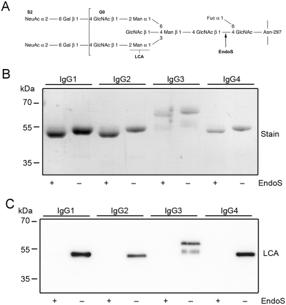
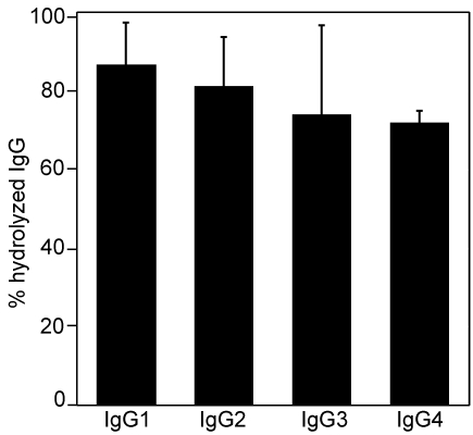
Background: The human pathogen Streptococcus pyogenes produces an endoglycosidase, EndoS that hydrolyzes the chitobiose core of the asparagine-linked glycan on the heavy chain of human IgG. IgG-binding to Fc gamma receptors (Fc gamma R) on leukocytes triggers effector functions including phagocytosis, oxidative burst and the release of inflammatory mediators. The interactions between Fc gamma R and the Fc domain of IgG depend on the IgG glycosylation state.

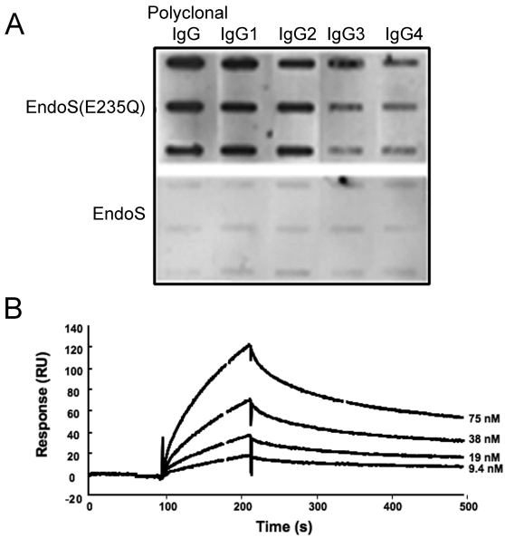
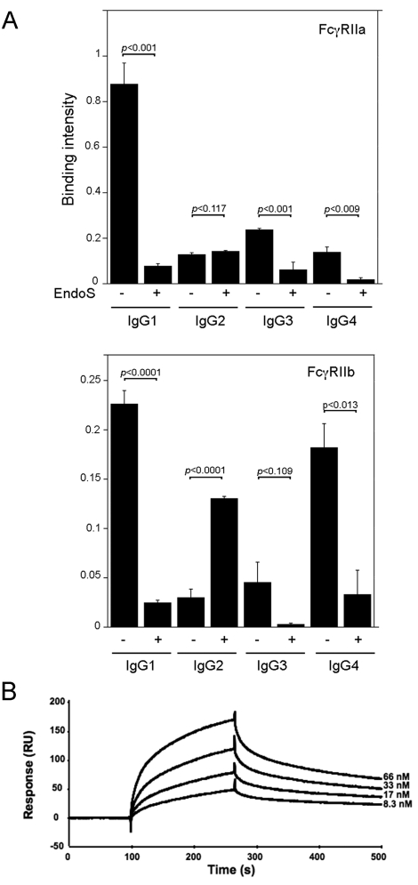
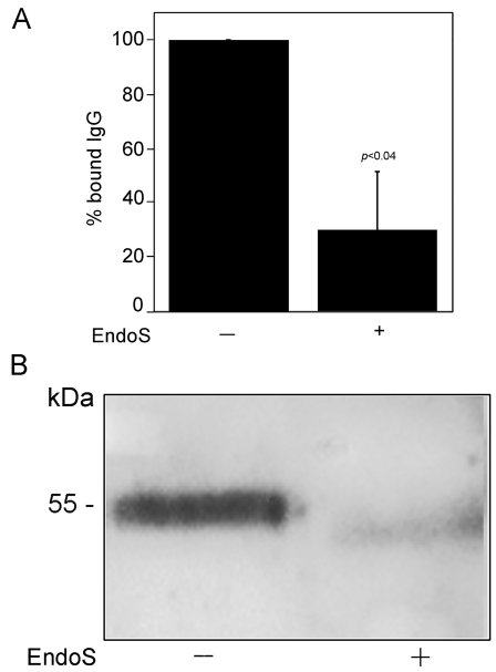
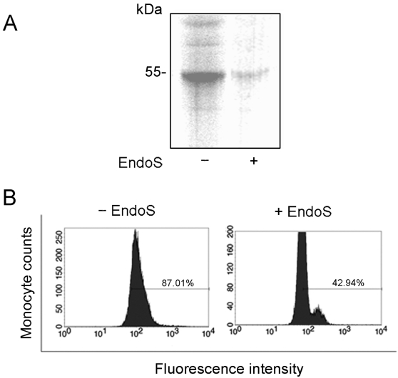
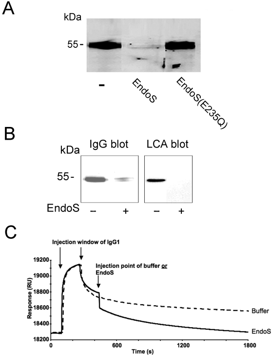
Methodology/principal findings: Here we show for the first time that EndoS hydrolyzes the heavy chain glycan of all four human IgG subclasses (IgG1-4), in purified form and in a plasma environment. An inactive form of EndoS, obtained by site-directed mutagenesis, binds IgG with high affinity, in contrast to wild type EndoS that only transiently interacts with IgG, as shown by Slot-blotting and surface plasmon resonance technology. Furthermore, EndoS hydrolysis of the IgG glycan influences the binding of IgG to immobilized soluble Fc gamma R and to an erythroleukemic cell line, K562, expressing Fc gamma RIIa. Incubation of whole blood with EndoS results in a dramatic decrease of IgG binding to activated monocytes as analyzed by flow cytometry. Moreover, the IgG bound to K562 cells dissociates when cells are treated with EndoS. Likewise, IgG bound to immobilized Fc gamma RIIa and subsequently treated with EndoS, dissociates from the receptor as analyzed by surface plasmon resonance and Western blot.

Conclusions/significance: We provide novel information about bacterial enzymatic modulation of the IgG/Fc gamma R interaction that emphasizes the importance of glycosylation for antibody effector functions. Moreover, EndoS could be used as a biochemical tool for specific IgG N-glycan hydrolysis and IgG purification/detection, or as a potential immunosuppressing agent for treatment of antibody-mediated pathological processes.


Conflict of interest statement

Competing Interests: A patent application on the in vitro use of EndoS has been filed with MA, AO, FN, and MC listed as inventors. The application is pending.

Figures

Figure 1
Figure 1. EndoS has glycosidase activity on all four human IgG subclasses.
Panel A. Glycan structure of human IgG. Glycan on the γ−chains of IgG attached to aspargine 297. GlcNAc, N-acetylglucosamine; Fuc, fucose; Man, mannose; Gal, galactose; NeuAc, sialic acid. Cleavage site for EndoS and recognation site for Lens culinaris agglutinin lectin (LCA) are indicated. Panel B. Purified IgG 1–4 incubated with EndoS and analyzed by SDS-PAGE and stained. Panel C. IgG 1–4 incubated with EndoS and analyzed using LCA lectin blot.
Figure 2
Figure 2. Hydrolysis of human IgG-subclasses by EndoS in plasma environment.
Human plasma treated with EndoS or PBS and followed by IgG glycan hydrolysis detection using LCA lectin ELISA of the purified IgG fraction. The results are presented as percent hydrolysis of each subclass compared to signals from untreated plasma. Means and standard deviations, indicated with error bars, were calculated from three independed experiments using blood from three different donors.
Figure 3
Figure 3. EndoS(E235Q) binds to all IgG-subclasses.
Panel A. Slot-blot representing the binding of EndoS and EndoS (E235Q) to each IgG subclass immobilized onto a nitrocellulose membrane in amounts: 3, 1.5 and 0.75 µg. The binding was detected using antiserum against EndoS. Panel B. Binding of EndoS (E235Q) to immobilized IgG-classes using BIAcore technology. The selected plot shows EndoS (E235Q) binding to IgG1 using hydrolysed IgG1 as a reference (bulk changes subtracted).
Figure 4
Figure 4. EndoS treatment of IgG subclasses inhibits binding of IgG to FcγRII.
Panel A. Binding of purified IgG subclasses, with or without EndoS treatment, to FcγRIIa and FcγRIIb immobilized to a microtitter plate. HRP-labeled protein G was used for detection of bound IgG subclasses. (−) indicates intact IgG and (+) EndoS hydrolysed IgG. Means, standard deviations (indicated with error bars), and p values (calculated using Student's t-test) were determined from three separate experiments. Panel B. Binding of IgG subclasses to immobilized receptors as visualized using BIAcore surface plasmon resonance. Plot shows a typical sensorgram, here the IgG1 binding to FcγRIIa. An empty flow cell is used as reference (subtracted).
Figure 5
Figure 5. EndoS treated IgG does not bind to FcγRIIa on K562 cells. Panel A.
The relative binding of radioactive IgG, With or without EndoS treatment, to K562 cells. The cells were incubated with 125iodine-labelled IgG (intact or EndoS-treated). The radioactivity of the washed cell pellets was detected. The binding of 125I- IgG (intact) to K562 cells, presented here as 100%, represents a specific IgG binding to K562 cells that could be inhibited by addition of cold IgG. Means, standard deviations (indicated with error bars), and p values (calculated using Student's t-test) were determined from three separate experiments. Panel B. K562 cells were incubated with human plasma treated with EndoS or PBS. The cells were resuspended in lysis buffer and analyzed by SDS-PAGE and Western blot using antiserum against human IgG.
Figure 6
Figure 6. EndoS treated IgG does not bind to monocytes. Panel A.
Monocytes were incubated with 125iodine-labelled IgG (intact or EndoS-treated). After incubation for 30 minutes at room temperature, the proteins from cell lysates, 10 µg total protein, were separated by 10% SDS-PAGE. The gel was dried and analyzed by phosphorimaging. Panel B. Flow cytometry analysis showing the decreased binding of IgG to activated monocytes in blood treated with EndoS. Human blood was treated with EndoS before addition of leukocyte activator fMLP. The IgG binding to monocytes was detected using mouse anti-human IgG and FITC-labelled goat anti-mouse IgG as a secondary antibody.
Figure 7
Figure 7. Dissociation of IgG from FcγRII upon treatment with EndoS. Panel A.
IgG bound to K562 cells dissociates from FcγRIIa upon incubation with EndoS but not with EndoS(E235Q). K562 cells were incubated with plasma and subsequently with EndoS, EndoS(E235Q) or PBS. Cell lysates, 10 µg total protein, were analysed for IgG by SDS-PAGE and blot using antiserum against human IgG. Panel B. IgG bound to monocytes dissociates from FcγRs after treatment with EndoS. Monocytes were incubated with plasma and later with EndoS or PBS. Resuspended cell pellets were analyzed for IgG by blot using antiserum against human IgG. The glycan of IgG was detected by blot and reactivity with LCA lectin. Panel C. A BIAcore setup showing EndoS affecting the IgG1 dissociation from an immobilized receptor FcγRIIa. In two parallel experiments, the injection of EndoS (black curve) is compared to the injection of buffer (broken line) at the same time-point during the dissociation phase of the IgG1- FcγRIIa interaction.

References

    1. Ravetch JV, Bolland S. IgG Fc receptors. Annu Rev Immunol. 2001;19:275–290. - PubMed
    1. Burton DR, Woof JM. Human antibody effector function. Adv Immunol. 1992;51:1–84. - PubMed
    1. Arnold JN, Wormald MR, Sim RB, Rudd PM, Dwek RA. The impact of glycosylation on the biological function and structure of human immunoglobulins. Annu Rev Immunol. 2007;25:21–50. - PubMed
    1. Jefferis R, Lund J, Pound JD. IgG-Fc-mediated effector functions: molecular definition of interaction sites for effector ligands and the role of glycosylation. Immunol Rev. 1998;163:59–76. - PubMed
    1. Dube R, Rook GA, Steele J, Brealey R, Dwek R, et al. Agalactosyl IgG in inflammatory bowel disease: correlation with C-reactive protein. Gut. 1990;31:431–434. - PMC - PubMed

Publication types