Glutamine synthetase gene expression during the regeneration of the annelid Enchytraeus japonensis
- PMID: 18183418
- PMCID: PMC2265772
- DOI: 10.1007/s00427-007-0198-4
Glutamine synthetase gene expression during the regeneration of the annelid Enchytraeus japonensis
Abstract
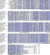
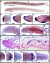
Enchytraeus japonensis is a highly regenerative oligochaete annelid that can regenerate a complete individual from a small body fragment in 4-5 days. In our previous study, we performed complementary deoxyribonucleic acid subtraction cloning to isolate genes that are upregulated during E. japonensis regeneration and identified glutamine synthetase (gs) as one of the most abundantly expressed genes during this process. In the present study, we show that the full-length sequence of E. japonensis glutamine synthetase (EjGS), which is the first reported annelid glutamine synthetase, is highly similar to other known class II glutamine synthetases. EjGS shows a 61-71% overall amino acid sequence identity with its counterparts in various other animal species, including Drosophila and mouse. We performed detailed expression analysis by in situ hybridization and reveal that strong gs expression occurs in the blastemal regions of regenerating E. japonensis soon after amputation. gs expression was detectable at the cell layer covering the wound and was found to persist in the epidermal cells during the formation and elongation of the blastema. Furthermore, in the elongated blastema, gs expression was detectable also in the presumptive regions of the brain, ventral nerve cord, and stomodeum. In the fully formed intact head, gs expression was also evident in the prostomium, brain, the anterior end of the ventral nerve cord, the epithelium of buccal and pharyngeal cavities, the pharyngeal pad, and in the esophageal appendages. In intact E. japonensis tails, gs expression was found in the growth zone in actively growing worms but not in full-grown individuals. In the nonblastemal regions of regenerating fragments and in intact worms, gs expression was also detected in the nephridia, chloragocytes, gut epithelium, epidermis, spermatids, and oocytes. These results suggest that EjGS may play roles in regeneration, nerve function, cell proliferation, nitrogenous waste excretion, macromolecule synthesis, and gametogenesis.
Figures


Similar articles
-
Molecular approach to annelid regeneration: cDNA subtraction cloning reveals various novel genes that are upregulated during the large-scale regeneration of the oligochaete, Enchytraeus japonensis.Dev Dyn. 2006 Aug;235(8):2051-70. doi: 10.1002/dvdy.20849. Dev Dyn. 2006. PMID: 16724321
-
Functional analysis of grimp, a novel gene required for mesodermal cell proliferation at an initial stage of regeneration in Enchytraeus japonensis (Enchytraeidae, Oligochaete).Int J Dev Biol. 2010;54(1):151-60. doi: 10.1387/ijdb.082790mt. Int J Dev Biol. 2010. PMID: 19876829
-
Morphallactic regeneration as revealed by region-specific gene expression in the digestive tract of Enchytraeus japonensis (Oligochaeta, Annelida).Dev Dyn. 2008 May;237(5):1284-94. doi: 10.1002/dvdy.21518. Dev Dyn. 2008. PMID: 18393309
-
Stem cell system in asexual and sexual reproduction of Enchytraeus japonensis (Oligochaeta, Annelida).Dev Growth Differ. 2010 Jan;52(1):43-55. doi: 10.1111/j.1440-169X.2009.01149.x. Epub 2009 Dec 20. Dev Growth Differ. 2010. PMID: 20039928 Review.
-
Molecular approach to echinoderm regeneration.Microsc Res Tech. 2001 Dec 15;55(6):474-85. doi: 10.1002/jemt.1192. Microsc Res Tech. 2001. PMID: 11782076 Review.
Cited by
-
Possible roles of glutamine synthetase in responding to environmental changes in a scleractinian coral.Mol Biol Rep. 2018 Dec;45(6):2115-2124. doi: 10.1007/s11033-018-4369-3. Epub 2018 Sep 10. Mol Biol Rep. 2018. PMID: 30203242
-
Molecular cloning and characterization of glutamine synthetase, a tegumental protein from Schistosoma japonicum.Parasitol Res. 2012 Dec;111(6):2367-76. doi: 10.1007/s00436-012-3092-6. Epub 2012 Sep 26. Parasitol Res. 2012. PMID: 23011789
-
What role do annelid neoblasts play? A comparison of the regeneration patterns in a neoblast-bearing and a neoblast-lacking enchytraeid oligochaete.PLoS One. 2012;7(5):e37319. doi: 10.1371/journal.pone.0037319. Epub 2012 May 16. PLoS One. 2012. PMID: 22615975 Free PMC article.
-
Expression level of glutamine synthetase is increased in hepatocellular carcinoma and liver tissue with cirrhosis and chronic hepatitis B.Hepatol Int. 2011 Jun;5(2):698-706. doi: 10.1007/s12072-010-9230-2. Epub 2010 Dec 28. Hepatol Int. 2011. PMID: 21484108 Free PMC article.
-
Injury-Induced Innate Immune Response During Segment Regeneration of the Earthworm, Eisenia andrei.Int J Mol Sci. 2021 Feb 27;22(5):2363. doi: 10.3390/ijms22052363. Int J Mol Sci. 2021. PMID: 33673408 Free PMC article.
References
-
- Abe S, Katagiri T, Saito-Hisaminato A, Usami S, Inoue Y, Tsunoda T, Nakamura Y. Identification of CRYM as a candidate responsible for nonsyndromic deafness, through cDNA microarray analysis of human cochlear and vestibular tissues. Am J Hum Genet. 2003;72:73–82. doi: 10.1086/345398. - DOI - PMC - PubMed
-
- Allodi S, Bressan CM, Carvalho SL, Cavalcante LA. Regionally specific distribution of the binding of anti-glutamine synthetase and anti-S100 antibodies and of Datura stramonium lectin in glial domains of the optic lobe of the giant prawn. Glia. 2006;53:612–620. doi: 10.1002/glia.20317. - DOI - PubMed
-
- Carlson BM. Development and regeneration, with special emphasis on the amphibian limb. In: Ferretti P, Géraudie J, editors. Cellular and molecular basis of regeneration: from invertebrates to Human. Chichester, NY: Wiley; 1998. pp. 411–450.
-
- Edwards CA, Bohlen PJ. Biology and ecology of earthworms. 3. London: Chapman & Hall; 1996.
Publication types
MeSH terms
Substances
LinkOut - more resources
Full Text Sources

