. 2008 Jan;6(1):e2.
doi: 10.1371/journal.pbio.0060002.

A mechanism regulating the onset of Sox2 expression in the embryonic neural plate

Affiliations

A mechanism regulating the onset of Sox2 expression in the embryonic neural plate

Costis Papanayotou et al. PLoS Biol. 2008 Jan.

Abstract

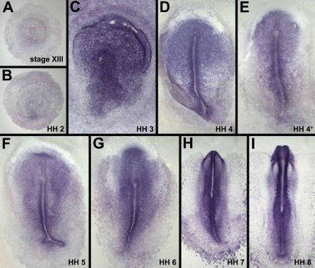
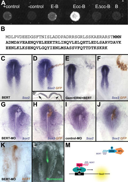
In vertebrate embryos, the earliest definitive marker for the neural plate, which will give rise to the entire central nervous system, is the transcription factor Sox2. Although some of the extracellular signals that regulate neural plate fate have been identified, we know very little about the mechanisms controlling Sox2 expression and thus neural plate identity. Here, we use electroporation for gain- and loss-of-function in the chick embryo, in combination with bimolecular fluorescence complementation, two-hybrid screens, chromatin immunoprecipitation, and reporter assays to study protein interactions that regulate expression of N2, the earliest enhancer of Sox2 to be activated and which directs expression to the largest part of the neural plate. We show that interactions between three coiled-coil domain proteins (ERNI, Geminin, and BERT), the heterochromatin proteins HP1alpha and HP1gamma acting as repressors, and the chromatin-remodeling enzyme Brm acting as activator control the N2 enhancer. We propose that this mechanism regulates the timing of Sox2 expression as part of the process of establishing neural plate identity.


Conflict of interest statement

Competing interests. The authors have declared that no competing interests exist.

Figures

Figure 1
Figure 1. HP1α Inhibits Sox2
(A, C, E, G, I, K, M, and Q) Embryos electroporated with BrmK755R (A), GFP (C), or HP1α (E) in the neural plate, ΔHP1α (G) or GFP (I) in the nonneural ectoderm, and ΔHP1α (K), GFP (M), and ΔHP1α together with BrmK755R (Q) in the extra-embryonic epiblast and stained for Sox2 (purple). (B, D, F, H, J, L, N, and R) Subsequent staining for GFP (brown) marks the electroporated cells in the same embryos. (O and P) Embryo electroporated with ΔHP1α in the extra-embryonic epiblast and stained for Brachyury (purple) (O). In (P), the same embryo is stained for GFP (brown), which marks the electroporated cells. (S) These results suggest that HP1α bound to Brm on the N2 enhancer inhibits expression of Sox2. In this and subsequent figures, the construct electroporated is indicated on the lower left, and the probes used for in situ hybridization and antibody staining are on the lower right of the panels.
Figure 2
Figure 2. Expression of HP1α during Normal Development
(A–C) Before and during gastrulation, HP1α is expressed throughout the embryo although its expression becomes gradually stronger in the prospective neural plate. (D–I) At the end of gastrulation (D), HP1α expression in the ectoderm becomes restricted in the prospective neural plate, where it gets stronger in subsequent stages (E–I) while it disappears from the nonneural ectoderm and the extra-embryonic epiblast. The number on each panel represents the embryonic stage according to Eyal-Giladi and Kochav [60] for pre-primitive streak stages (in Roman numerals), and Hamburger and Hamilton [7] for later stages (Arabic numerals).
Figure 3
Figure 3. Expression of Chick Geminin during Normal Development and Its Regulation by FGF
(A–G) Geminin is expressed in the embryonic epiblast from pre-primitive streak stages (A). As the embryo develops, its expression becomes restricted to the neural plate, where it intensifies (B–G). (H) An FGF-soaked bead up-regulates Geminin in the extra-embryonic epiblast (arrow); a control bead has been grafted on the contralateral side (arrowhead). Embryo stages as for Figure 2.
Figure 4
Figure 4. Geminin Induces Sox2
(A–F) Embryos electroporated with Geminin (Gem) in the nonneural ectoderm (A) and Geminin (C) or Geminin together with BrmK755R (BrmDN) (E) in the extra-embryonic epiblast and stained for Sox2 (purple). Subsequent staining for GFP (brown) marks the electroporated cells in the same embryos (B), (D), and (F). (G) We propose that Geminin displaces HP1α from its binding site on Brm, releasing its inhibition on Sox2 expression.
Figure 5
Figure 5. ERNI Blocks Induction of Sox2 by Geminin
(A and B) Embryo electroporated with Geminin and ERNI in the extraembryonic epiblast and stained for Sox2 (A) and GFP (B) to mark the electroporated cells. (C) We propose that ERNI inhibits the induction of Sox2 by Geminin.
Figure 6
Figure 6. Mutated Forms of ERNI Induce Sox2
(A, C, E, and G) Embryos electroporated with the isolated coiled-coil domain of ERNI (ERNIcc) (A), ERNI (C), GFP (E), or ERNIY228F (G) in the nonneural ectoderm and stained for Sox2 (purple). (B, D, F, and H) Subsequent staining for GFP (brown) marks the electroporated cells in the same embryos.
Figure 7
Figure 7. ERNI Inhibits Sox2 through HP1γ
(A) A two-hybrid screen reveals that HP1γ interacts with the C-terminus of ERNI: ++, +, or − indicates growth induced by interaction of ENS-1/ERNI (E) with HP1γ/CHCB2 or with itself. Partially deleted or HP1-box–mutated forms are indicated. HP1γ-CSD: carboxy-terminal 87 aa of HP1γ containing the chromoshadow domain (CSD). (B) Partial amino acid sequence of ENS-1/ERNI: the pentapeptide PXVXL, necessary for specific interaction with the CSD of HP1γ/CHCB2, is in bold. (C) Embryo electroporated with Geminin, ERNI, and ΔHP1γ in the extra-embryonic epiblast (G) and stained for Sox2 (purple). (D) The same embryo after staining for GFP (brown) to mark electroporated cells. (E) We propose that ERNI inhibits Sox2 expression by recruiting HP1γ to the N2 enhancer.
Figure 8
Figure 8. Expression of HP1γ during Normal Development
(A) Before primitive streak formation, HP1γ is expressed in the extra-embryonic epiblast. (B–D) When the primitive streak forms, HP1γ expression appears in the embryonic epiblast where it gradually becomes stronger in the prospective neural plate. (E–I) At the end of gastrulation (E), its expression in the ectoderm becomes restricted in the prospective neural plate, where it gets stronger at subsequent stages (F–I), while it disappears from the nonneural ectoderm and extra-embryonic epiblast.
Figure 9
Figure 9. BERT Releases Geminin from the Inhibition of ERNI
(A) Representative colonies from the two-hybrid screen (left to right): positive (+) control, negative (−) control, ERNI+BERT (E-B), ERNIcc+BERT (Ecc-B), ERNI without coiled-coil domain + BERT (EΔcc-B), and BERT alone (B). (B) Amino acid sequence of BERT. The sequence originally isolated is shown in bold; the rest encodes a putative upstream exon. (C–F) Embryos electroporated with BERT (C) in the nonneural ectoderm or Geminin, ERNI, and BERT (E) in the extra-embryonic epiblast and stained for Sox2 (purple). The same embryos after staining for GFP (brown) to mark electroporated cells (D) and (F). A section of the embryo in (D) reveals that the induced epiblast acquires a neural plate–like morphology (D′). (G–J) Embryos electroporated with a MO against BERT (G) or a control MO (I) in the prospective neural plate and stained for Sox2 (purple). In (H) and (J), the same embryos are stained with anti-fluorescein to detect the MO (brown). (K and L) Embryo electroporated with the MO against BERT in the neural plate and immunostained for BERT protein (brown, [K]). (L) The same embryo under fluorescence to show the cells electroporated with the fluorescein-labeled MO. (M) We propose that BERT disrupts the interaction between Geminin and ERNI, displacing HP1γ from the N2 enhancer and thus allowing Geminin/Brahma to induce Sox2 expression.
Figure 10
Figure 10. Expression of BERT during Normal Development
BERT is expressed ubiquitously in the chick embryo albeit at low levels. At the end of gastrulation (A), its expression is up-regulated in the prospective neural plate, where it becomes stronger during subsequent stages (B–D).
Figure 11
Figure 11. Direct Interactions between the Coiled-Coil Proteins
(A–C) Example of a positive interaction between BERT and itself (homodimerization). B(C): BERT-Venus(C); B(N), BERT-Venus(N). (D and F) Example of a negative control, BERT, and another coiled-coil protein, E2F3. D(C), E2F5-Venus(C). (A) and (D) Phase contrast, (B) and (E) fluorescence, (C) and (F) merged phase contrast and fluorescence images.
Figure 12
Figure 12. BiFCo Competition Assays
(A and B) BERT disrupts Geminin-Venus(N)::ERNI-Venus(C) heterodimers (G(N)-E(C)). B, BERT. (D and E) BERT-Venus(C) disrupts Geminin-Venus(N)::ERNI heterodimers through its association with Geminin-Venus(N). B(C), Bert-Venus(C); E, ERNI; G(N), Geminin-Venus(N). (G and H) BERT-Venus(C) disrupts Geminin::ERNI-Venus(N) heterodimers through its association with ERNI-Venus(N). B(C), BERT-Venus(C); E(N), ERNI-Venus(N); G, Geminin. (C), (F), and (I) are the respective controls using the noninteracting Dlx5 protein as competitor. D(C), Dlx5-Venus(C)
Figure 13
Figure 13. Interaction of Geminin, ERNI, and BERT on the N2 Enhancer
(A–D) Electroporation of GFP with N2-TK-LacZ does not activate the reporter (A), whereas electroporation of Geminin+ERNI+BERT with N2-TK-LacZ does ([C]; LacZ in blue). In (B) and (D), the same embryos are stained for GFP to mark the electroporated cells. (E) ChIP assay on chromatin from E7.5 mouse neural plate, demonstrating a direct interaction between Geminin and the N2 enhancer. ab, antibody.

Similar articles

Cited by

References

    1. Avilion AA, Nicolis SK, Pevny LH, Perez L, Vivian N, et al. Multipotent cell lineages in early mouse development depend on SOX2 function. Genes Dev. 2003;17:126–140. - PMC - PubMed
    1. Bylund M, Andersson E, Novitch BG, Muhr J. Vertebrate neurogenesis is counteracted by Sox1–3 activity. Nat Neurosci. 2003;6:1162–1168. - PubMed
    1. Graham V, Khudyakov J, Ellis P, Pevny L. SOX2 functions to maintain neural progenitor identity. Neuron. 2003;39:749–765. - PubMed
    1. Ellis P, Fagan BM, Magness ST, Hutton S, Taranova O, et al. SOX2, a persistent marker for multipotential neural stem cells derived from embryonic stem cells, the embryo or the adult. Dev Neurosci. 2004;26:148–165. - PubMed
    1. Donner AL, Episkopou V, Maas RL. Sox2 and Pou2f1 interact to control lens and olfactory placode development. Dev Biol. 2007;303:784–799. - PMC - PubMed

Publication types

MeSH terms

Associated data