. 2008 Jan 10;57(1):41-55.
doi: 10.1016/j.neuron.2007.11.018.

Members of the miRNA-200 family regulate olfactory neurogenesis

Affiliations

Members of the miRNA-200 family regulate olfactory neurogenesis

Philip S Choi et al. Neuron. .

Abstract

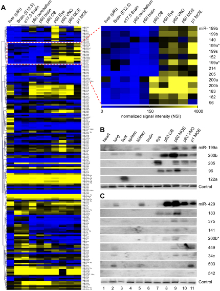
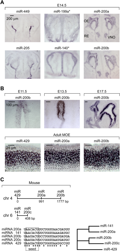
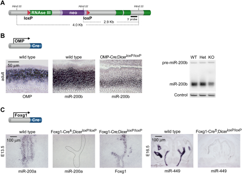
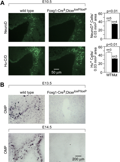
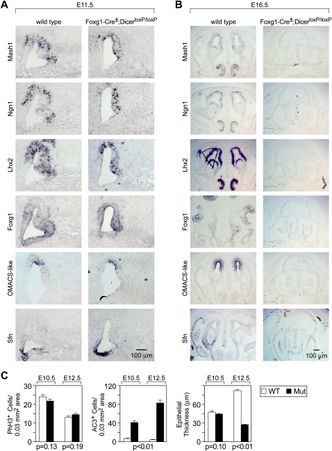
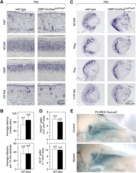
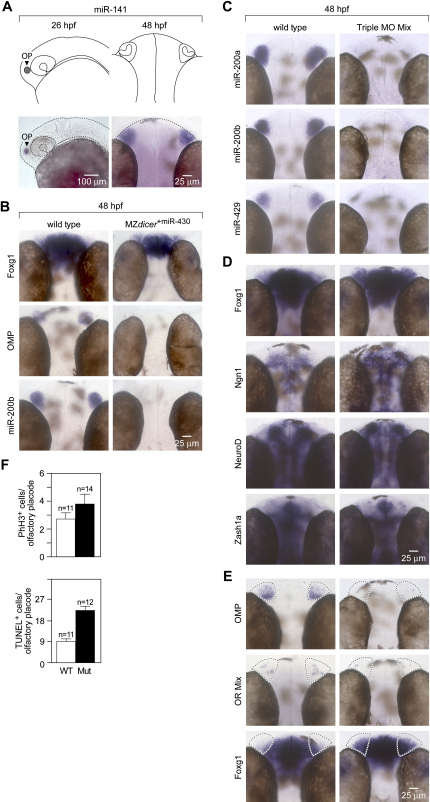
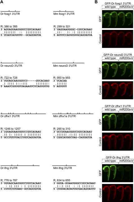
MicroRNAs (miRNAs) are highly expressed in vertebrate neural tissues, but the contribution of specific miRNAs to the development and function of different neuronal populations is still largely unknown. We report that miRNAs are required for terminal differentiation of olfactory precursors in both mouse and zebrafish but are dispensable for proper function of mature olfactory neurons. The repertoire of miRNAs expressed in olfactory tissues contains over 100 distinct miRNAs. A subset, including the miR-200 family, shows high olfactory enrichment and expression patterns consistent with a role during olfactory neurogenesis. Loss of function of the miR-200 family phenocopies the terminal differentiation defect observed in absence of all miRNA activity in olfactory progenitors. Our data support the notion that vertebrate tissue differentiation is controlled by conserved subsets of organ-specific miRNAs in both mouse and zebrafish and provide insights into control mechanisms underlying olfactory differentiation in vertebrates.


Figures

Figure 1
Figure 1
Identification of Olfactory miRNAs by Microarray and Cloning Approaches (A) Hierarchical clustering of miRNA expression profiles from several tissues using microRNA microarrays (Miska et al., 2004). The cluster of miRNAs with predicted enrichment in olfactory tissues is highlighted (right panel). Blue color indicates weak hybridization signals, and yellow indicates strong hybridization signals. miRNAs are considered present in a given tissue if they display a normalized signal intensity (NSI) ≥ 100. (B and C) Validation by northern blot analysis of miRNAs identified by microarray and cloning strategies. All tissue samples originate from adult mice (P60), excluding rat VNO (P60) and rat MOE (P1). miR-122a, known to be exclusively expressed in liver tissue, is used as a positive control. U6 snRNA serves as a loading control.
Figure 2
Figure 2
Expression Patterns of Olfactory miRNAs Analyzed by Locked Nucleic Acid-Based In Situ Hybridization (A) Three basic patterns of miRNA expression were identified during embryonic MOE development. Left: the expression of miR-34b, 34c, 139, 205, and 449 is restricted to the respiratory epithelium. Middle: the expression of miR-125b, 140, 199a, 199a, and 199b is restricted to the mesenchyme underlying or cartilage surrounding the MOE. Right: the expression of miR-96, 141, 182, 183, 200a, 200b, 191, and 429 is strongest in the MOE and VNO neuroepithelium, with reduced levels in the respiratory epithelium. OE, olfactory epithelium; RE, respiratory epithelium; VNO, vomeronasal organ. (B) Developmental time course analysis of miR-200 family member expression. (C) Genomic organization of mouse miR-200 family members.
Figure 3
Figure 3
Conditional Ablation of Dicer in Mature Olfactory Neurons and Olfactory Progenitors (A) Schematic diagram of the Dicer conditional targeting construct used in this study (Harfe et al., 2005). (B) Cross of OMP-Cre and DicerloxP/loxP transgenic lines. miR-200b and OMP expression overlaps in mature neurons (left and center panels). Mature miR-200b expression is abolished in OMP-expressing cells of OMP-Cre; DicerloxP/loxP mice but remains in OMP-negative, immature neurons and progenitor cells located in the basal MOE (right panel). Broken black line indicates the basal lamina of the MOE. Northern blot analysis confirms the reduction in miR-200b expression (right blot). (C) Cross of Foxg1-Cre and DicerloxP/loxP transgenic lines. Tissues derived from the olfactory placodes of Foxg1-Cre+/; DicerloxP/loxP tissues were analyzed for expression of mature miR-200a and miR-449 expression. Expression of Foxg1 in adjacent sections was used to demonstrate that MOE and respiratory epithelial tissue is still present in these mutants.
Figure 4
Figure 4
Olfactory Precursor Cells of Foxg1-Cre+/; DicerloxP/loxP Mutants Display Normal Specification but Do Not Fully Differentiate (A) Number of differentiating and postmitotic cells in olfactory placodes was quantified by neuroD (mean ± SEM, WT 41.71 ± 2.10, n = 5; mutant 34.33 ± 1.60, n = 4, p < 0.01, Student's t test) and Hu-C/D (mean ± SEM, WT 45.82 ± 2.57, n = 3; mutant 31.32 ± 2.09, n = 3, p < 0.01, Student's t test) expression, respectively, in Foxg1-Cre+/; DicerloxP/loxP and control E10.5 embryos. Only moderate reduction in the number of precursor cells and postmitotic neurons is observed in the mutant at this stage. Cell counts were derived from sections spanning the entire nasal pit of several animals per genotype and normalized to 0.03 mm2; the average MOE in a given section. (B) In situ hybridization on E13.5 olfactory epithelium fails to detect OMP expression in Foxg1-Cre+/; DicerloxP/loxP olfactory placodes, suggesting the failure of olfactory terminal differentiation in the absence of Dicer function.
Figure 5
Figure 5
Olfactory Precursor Cells of Foxg1-Cre+/; DicerloxP/loxP Mutants Display Normal Patterning but Do Not Fully Differentiate (A) Foxg1-Cre+/; DicerloxP/loxP olfactory placodes at E11.5 were assayed for expression of markers that distinguish olfactory progenitor cells (Mash1, Ngn1, Lhx2, and Foxg1), MOE zonal patterning (OMACS-like), and respiratory epithelium (Sfn). Expression of these genes suggests normal gross patterning. (B) Cells of the olfactory neuronal cell lineages are lost, while nonneuronal cell lineages are maintained in Foxg1-Cre+/;DicerloxP/loxP mutant MOE by E16.5. Expression of markers that distinguish olfactory neurogenesis (Mash1, Ngn1, Lhx2, and Foxg1) and zonal patterning (OMACS-like) cannot be detected in Foxg1-Cre+/;DicerloxP/loxP mutant MOE at E16.5. By contrast, expression of respiratory epithelium (Sfn) persists in mutant MOE. In addition, the normally convoluted structure of the MOE is reduced to a simple epithelium comprised solely of nonneural respiratory epithelium. (C) Quantification of phospho-histone H3 and active caspase-3 immunoreactive cells in embryonic MOE of Foxg1-Cre+/; DicerloxP/loxP mutants and controls at E10.5 (mean ± SEM, WT 23.95 ± 1.06, n = 3; mutant 21.61 ± 1.09, n = 3, p = 0.13, Student's t test) and E12.5 (mean ± SEM, WT 13.02 ± 0.76 cells, n = 3; mutant 14.49 ± 0.77 cells, n = 3, p = 0.19, Student's t test) and active caspase-3 at E10.5 (mean ± SEM, WT 7.76 ± 1.44, n = 3; mutant 41.97 ± 3.31, n = 3, p < 0.01, Student's t test) and E12.5 (mean ± SEM, WT 5.18 ± 0.54, n = 3; mutant 83.42 ± 5.54, n = 3, p < 0.01, Student's t test) indicate that loss of Dicer function results in increased cellular apoptosis and unchanged cellular proliferation in the olfactory epithelium.
Figure 6
Figure 6
Ablation of Dicer Function in Mature Olfactory Sensory Neurons Does Not Cause Any Apparent Molecular or Behavioral Defects (A) OMP-Cre; DicerloxP/loxP adult MOE (P60) showed normal expression of molecular markers that identifies olfactory progenitor proliferation (Ki67), olfactory neuron differentiation (NCAM), and mature olfactory neurons (OMP and olfactory receptors). (B) Time required to discover a hidden cookie (latency) by OMP-Cre; DicerloxP/loxP mutant mice and control animals (mean ± SEM, WT 66.14 ± 27.91 s; mutant 88.63 ± 19.83 s; p = 0.53, Students t test) was statistically indistinguishable. Similarly, quantification of resident average attack frequency in a resident-intruder assay designed to test VNO function in OMP-Cre; DicerloxP/loxP mutants and control animals (mean ± SEM, WT 35.6 ± 13.65 s; mutant 35.75 ± 15.93 s; p = 0.99, Student's t test) showed no significant difference. (C) OMP-Cre; DicerloxP/loxP adult MOE (P60) showed normal expression of molecular markers for vomeronasal neuronal differentiation (NCAM), zonal patterning (G protein subunits) and mature function (V1 receptors). (D) Quantification of phospho-histone H3 immunoreactive cells (mean ± SEM, WT 5.79 ± 0.50, n = 3; mutant 5.05 ± 0.37, n = 3, p = 0.24, Student's t test) and active caspase-3 immunoreactive cells (mean ± SEM, WT 12.19 ± 0.77, n = 3; mutant 11.76 ± 0.74, n = 3, p = 0.69, Student's t test) in adult MOE of OMP-Cre; DicerloxP/loxP mutants and controls reveals no statistically significant differences in proliferation or apoptosis rates. (E) OMP-Cre; DicerloxP/loxP; P2-IRES-TauLacZ triple-transgenic mice (P45) showed normal expression and axon targeting of LacZ in P2-expressing olfactory neurons.
Figure 7
Figure 7
Zebrafish miR-200 Family Members Are Required for Terminal Differentiation of Olfactory Progenitor Cells (A) Schematic diagram of the zebrafish olfactory placode and olfactory organ at 26 hpf and 48 hpf, respectively, and corresponding expression pattern of miR-141, a member of the miR-200 family. (B) MZdicer embryos were injected with miR-430 (MZdicer+miR-430) to substantially rescue general neuronal and other phenotypic defects observed in MZdicer mutants by supplying the critical miRNA expressed during the earliest stages of development (Giraldez et al., 2005). MZdicer+miR-430 embryos assayed for expression of olfactory progenitor (foxg1), mature neuron (OMP), and miRNA (miR-200b) markers show impaired terminal differentiation of olfactory progenitors. (C) In situ hybridization staining of 48 hpf embryos for expression of miR-200a, miR-200b, and miR-420 that were injected at the one-cell stage with a combination of miR-141 MO, miR-200b MO, and miR-429 MO (4 ng each; Triple MO Mix) show complete loss of miR-200 family expression. (D) Wild-type and fish injected with various morpholinos at 48 hpf are morphologically indistinguishable from each other with the exception of expanded Foxg1 expression (see panel [E]). (E) Triple MO morphants injected at the one-cell stage and assayed for expression of olfactory progenitor marker (foxg1) and mature neuronal markers (OMP and an olfactory receptor mix) at 48 hpf show impaired terminal differentiation of olfactory progenitors. (F) Quantification of phospho-histone H3 immunoreactive cells (mean ± SEM, WT 2.55 ± 0.45, n = 11; morphant 3.57 ± 0.67, n = 14, p = 0.24, Student's t test) and TUNEL immunoreactive cells (mean ± SEM, WT 12.55 ± 1.46, n = 11; mutant 30.67 ± 2.59, n = 12, p < 0.01, Student's t test) in 72 hpf Triple MO morphant olfactory epithelia and controls reveals a statistically significant difference in apoptosis, but not proliferation.
Figure 8
Figure 8
miR-200 Target Validation (A) Comparison of conserved miR-200 sites in the 3′UTRs of select miR-200 predicted targets in mouse and zebrafish suggest that miR-200 family members may be sufficient to negatively regulate zfhx1, foxg1, and lfng and may help to downregulate neuroD. Vertical ticks on schematic drawings indicate a predicted miR-200 site, and the alignments correspond to the strongest miR-200 site produced by the miRanda algorithm (Enright et al., 2003). (B) GFP reporters fused upstream of full-length zebrafish 3′UTRs corresponding to putative targets containing predicted miR-200 binding sites were coinjected with control DsRed mRNA into wild-type zebrafish embryos at the one-cell stage either in the absence or presence of synthetic miR-200a/miR-200b RNA duplex. Fluorescent microscopy shows GFP reporter expression (green) and control DsRed expression (red) at 25–30 hpf, indicating that miR-200 family members are sufficient to downregulate zebrafish zfhx1 and lfng.

Similar articles

Cited by

References

    1. Andl T., Murchison E.P., Liu F., Zhang Y., Yunta-Gonzalez M., Tobias J.W., Andl C.D., Seykora J.T., Hannon G.J., Millar S.E. The miRNA-processing enzyme Dicer is essential for the morphogenesis and maintenance of hair follicles. Curr. Biol. 2006;16:1041–1049. - PMC - PubMed
    1. Bartel D.P. MicroRNAs: genomics, biogenesis, mechanism, and function. Cell. 2004;116:281–297. - PubMed
    1. Beites C.L., Kawauchi S., Crocker C.E., Calof A.L. Identification and molecular regulation of neural stem cells in the olfactory epithelium. Exp. Cell Res. 2005;306:309–316. - PubMed
    1. Belluscio L., Koentges G., Axel R., Dulac C. A map of pheromone receptor activation in the mammalian brain. Cell. 1999;97:209–220. - PubMed
    1. Bernstein E., Caudy A.A., Hammond S.M., Hannon G.J. Role for a bidentate ribonuclease in the initiation step of RNA interference. Nature. 2001;409:363–366. - PubMed

Publication types