. 2008 Jan 10;57(1):69-79.
doi: 10.1016/j.neuron.2007.10.041.

Cell-nonautonomous function of ceramidase in photoreceptor homeostasis

Affiliations

Cell-nonautonomous function of ceramidase in photoreceptor homeostasis

Jairaj K Acharya et al. Neuron. .

Abstract

Neutral ceramidase, a key enzyme of sphingolipid metabolism, hydrolyzes ceramide to sphingosine. These sphingolipids are critical structural components of cell membranes and act as second messengers in diverse signal transduction cascades. Here, we have isolated and characterized functional null mutants of Drosophila ceramidase. We show that secreted ceramidase functions in a cell-nonautonomous manner to maintain photoreceptor homeostasis. In the absence of ceramidase, photoreceptors degenerate in a light-dependent manner, are defective in normal endocytic turnover of rhodopsin, and do not respond to light stimulus. Consistent with a cell-nonautonomous function, overexpression of ceramidase in tissues distant from photoreceptors suppresses photoreceptor degeneration in an arrestin mutant and facilitates membrane turnover in a rhodopsin null mutant. Furthermore, our results show that secreted ceramidase is internalized and localizes to endosomes. Our findings establish a role for a secreted sphingolipid enzyme in the regulation of photoreceptor structure and function.


Figures

Figure 1
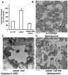
Figure 1. CDase protein is still present in cdase1 mutant clones
(A). cdase1 mosaic eye is generated using FRT-FLP recombination in conjunction with a cell lethal mutation on the wild type chromosome. This resulted in an almost entire eye derived from mutant cells except for small red spots (marked by arrow) that are heterozygous for CDase. (B). Transmission electron micrograph showing 15 day old w1118 and cdase1 clone of photoreceptors. Mutant photoreceptors generated by mitotic recombination in heterozygotes do not show signs of degeneration and are similar in structure and organization to wild type photoreceptors. (C). Eye imaginal discs from w1118 and cdase1 mutant clones are stained with anti-CDase antibody. cdase1 mutant clones are immunopositive for CDase protein while functional null cdase1 mutant (described later) does not show antibody reactivity. (D). Western analysis of retinal extracts from w1118, cdase1 and crumbs mutant clones reveals that CDase protein can be detected in cdase extract (upper panel) whereas crumbs extract generated similarly does not contain Crumbs protein (lower panel). cdase1 null mutant described later does not have any CDase protein.
Figure 2
Figure 2. cdase1 functional null mutant is devoid of CDase protein and activity
(A). Western blot analysis of 1, 5 and 10 head extracts from w1118 and cdase1 null reveals that no CDase protein is detected in mutant lanes. The blot is probed for tubulin as a loading control. (B). Neutral CDase activity is carried out using [1-14C] Palmitoyl sphingosine as the substrate and measuring radioactivity in the released fatty acid. While control w1118 extract shows good CDase activity, cdase1 null extract does not show any activity.
Figure 3
Figure 3. cdase1 functional null mutant photoreceptors undergo light-dependent degeneration
(A). TEM of five-day old photoreceptors of w1118, cdase1 null, cdase1 null rescue flies (with genomic CDase on second chromosome) reared in light and cdase1 reared in dark. cdase1 null photoreceptors exposed to light show degeneration accompanied by loss of rhabdomeres and vacuolation of cells. Photoreceptor degeneration can be completely rescued in mutant flies expressing a genomic copy of CDase. cdase1 mutant flies raised in the dark do not show signs of degeneration seen in corresponding light exposed flies. Insets show a low magnification view of the photoreceptors of the corresponding genotype. (B). CDase protein stains the rhabdomeres and sub rhabdomeric region (marked by arrowheads) in wild type photoreceptors. Thin sections of w1118 photoreceptors are double stained for Rhodopsin and CDase proteins with respective antibodies. In wild type photoreceptors, CDase can be colocalized with Rhodopsin as shown in the overlay.
Figure 4
Figure 4. CDase expressed in fat body reaches eye discs, is active, suppresses degeneration in arr23 and promotes membrane turnover in ninaEI17
(A). CDase is over-expressed in w1118 using a fat body Gal4 driver. Crumbs protein driven similarly in the fat body is used as control. The top panel shows overexpression of CDase (upper right panel) and Crumbs (upper left panel) in extracts (10 μg protein) prepared from dissected fat bodies. Retina are also isolated from these flies and extracts (10 μg protein) are subjected to Western analysis. Retinal extracts show significant accumulation of CDase (lower right panel) but not Crumbs (lower left panel). (B). CDase is over-expressed using fat body Gal4 driver in cdase1 background. Eye discs are isolated from these flies (cdase1 null and cdase1 null expressing CDase) and stained with a monoclonal antibody to CDase. These discs show good staining for CDase compared to mutant discs that do not express fat body driven CDase. Inset shows that in addition to staining the cytoplasm, CDase staining is observed in punctate dots inside the cells. (C). CDase expressed in the fat body is active in retina. Retinae are isolated from cdase1 null mutants and cdase1 null expressing fat body driven CDase. Neutral CDase activity is carried out using radiolabeled palmitoyl sphingosine as substrate and measuring the radioactivity in the released fatty acid. While cdase1 null retina show no activity, retina from cdase1 null expressing CDase in fat body show high activity. (D). The top panel shows photoreceptors of arr23 and arr23 expressing fat body driven CDase. CDase expression suppresses degeneration seen in arr23. The bottom panel shows photoreceptors of ninaEI17 and ninaEI17 expressing fat body driven CDase. Expression of CDase facilitates turnover of involuting rhabdomeric membranes (marked by arrows) in the ninaEI17 null mutant.
Figure 5
Figure 5. cdase1 null shows increased ceramide levels that decrease upon overexpression of CDase. Sphingosine is not able to rescue photoreceptor degeneration in cdase1 null mutant
(A). Estimation of ceramide levels in control, cdase1 null and cdase1 null expressing fat body driven CDase by mass spectrometry. Identification of ceramide molecular species containing tetradecasphingenine was performed by negative-ion ESI/MS/MS. Comparison of total ceramide shows that mutant flies have about 2 fold more ceramide than control. Expression of CDase in the fat body in the mutant background results in significant decrease in ceramide levels. (B). Photoreceptors of w1118 flies fed sphingosine. Control w1118 flies were raised in food supplemented with N-acetyl-D-erythro sphingosine (14 carbon long chain base, native to Drosophila) at concentrations that rescued lace lethality. The eclosed flies were aged for seven days and their photoreceptors were observed by TEM. Rhabdomere architecture is not significantly altered in sphingosine fed wild type flies. (C). Photoreceptors of cdase1 null aged for seven days show extensive degeneration. (D). Photoreceptors of cdase1 null fed sphingosine. Sphingosine does not rescue photoreceptor degeneration in cdase1 null flies.
Figure 6
Figure 6. Extra-cellular CDase can bind ceramide on target cells, is internalized and internalized CDase can localize to the endocytic compartment
(A). Western analysis of RNAi mediated knock down of CDase in Drosophila S2 cells. The blot shows efficient reduction in both intra and extra-cellular CDase upon treatment with dsRNA for 48 h. CDase present in extra-cellular media is 7-8 fold more than that detected intra-cellularly. However since same amount of total protein was loaded in each lane and since extra-cellular media contains serum proteins, the blot does not reflect the actual distribution of CDase. Tubulin serves as a loading control for the blot. (B). Cells treated with dsRNA as described in panel A, are incubated with C12 NBD-ceramide. After incorporation of the fluorescent analog, cells are incubated with either V5 tagged partially purified CDase or buffer. After incubation, lipids are extracted and separated by TLC. 40% more NBD-ceramide is hydrolyzed to the product in cells that receive tagged CDase (NBD-Cer + CDase) compared to control cells (NBD-Cer no CDase). The right most TLC lane shows that tagged CDase used above is active in vitro in converting NBD-Cer to fatty acid. (C). Immunofluorescence analysis of S2 cells incubated with V5 tagged CDase shows that it can be internalized. When stained with an antibody to V5, CDase can be visualized in punctate dots within the cells. (D). Internalized CDase co-localizes with endocytic marker, Rab 11. S2 cells incubated with V5 tagged CDase are double stained with antibodies to Drosophila Rab11 and V5. DAPI is used to stain the nucleus. The overlay shows CDase localizes to structures that are positive for Rab11. (E). Internalized CDase does not colocalize with Lava Lamp, a Golgi marker.
Figure 7
Figure 7. cdase1 null is not defective in forward transport of Rh1 but defective in Arr-Rh1 interaction. cdase1 null does not respond to light
(A). Western analysis of Rh1 in light and dark raised w1118 (C) and cdase1 (M) retinal extracts. Rh1 level progressively decreases in light exposed mutant photoreceptors while dark raised flies do not show decrease in Rh1 level. Rh1 is in the fully mature 34 kd form in the mutant photoreceptors. The blot is probed with an antibody to Inositol polyphosphate 1-phosphatase (IPP) as loading control. (B). Biochemical analysis of endocytic turnover of Rh1-Arr complexes in photoreceptors. The panel shows Arr2 and Rh1 blots. In wild type extracts, Arr2 can be pelleted with Rh1 in blue light and released efficiently to the supernatant in blue followed by orange light regimen. cdase1 shows inefficient release of Arr2 under similar conditions and this defect is rescued by the introduction of a wild type copy of CDase. P and S represent pellet and supernatant fractions respectively. (C). Quantification of Arrestin2 bound in control, cdase1 null and rescue flies. The graph shows that 27% more of the total Arr2 remains bound to Rh1 compared to control flies while it is comparable to wild type in the rescued flies. (D). In wild type extracts, upon exposure to any light, most of Arr1 is in the pellet while in cdase1 null, a significant portion of it is in the supernatant. This defect is corrected in rescued flies. (E). Electroretinogram (ERG) recordings from w1118, cdase1 null, and Cdase; cdase1 null flies. Dark-raised flies were exposed to two consecutive 10-second pulses of white light (400-700 nm, 6600 lux), as indicated beneath ERG recordings. Shown are representative ERG recordings from at least 7 flies examined for each genotype. cdase1 null flies displayed no light responsiveness at all light intensities (0.6 to 6600 lux) tested. As shown, CDase; cdase1 null flies exhibited complete rescue of light responsiveness with signals indistinguishable from wild type.
Figure 8
Figure 8. cdase1 is defective in apoptosis and photoreceptor degeneration in cdase1 can be rescued by expression of the anti apoptotic protein, p35
(A). The top panel shows untreated eye discs from w1118 and cdase1 while the bottom panel shows eye discs that have been irradiated with X-rays, allowed to recover for 5h and stained with anti-Caspase antibody. Mutants reveal an increase in basal as well as damage-induced apoptosis as seen by the increased Caspase staining in the discs. (B). Expression of GMR-p35 in photoreceptors rescues degeneration in cdase1. TEMs of five-day old photoreceptors are shown. Mutant photoreceptors show loss of rhabdomeres and vacuolation, these defects are not seen in mutant photoreceptors expressing p35.

References

    1. Acharya U, Edwards MB, Jorquera RA, Silva H, Nagashima K, Labarca P, Acharya JK. Drosophila melanogaster Scramblases modulate synaptic transmission. J Cell Biol. 2006;173:69–82. - PMC - PubMed
    1. Acharya U, Patel S, Koundakjian E, Nagashima K, Han X, Acharya JK. Modulating sphingolipid biosynthetic pathway rescues photoreceptor degeneration. Science. 2003;299:1740–1743. - PubMed
    1. Acharya U, Mowen MB, Nagashima K, Acharya JK. Ceramidase expression facilitates membrane turnover and endocytosis of rhodopsin in photoreceptors. Proc Natl Acad Sci USA. 2004;101:1922–1926. - PMC - PubMed
    1. Acharya U, Acharya JK. Enzymes of sphingolipid metabolism in Drosophila melanogaster. Cell Mol Life Sci. 2005;62:128–142. - PMC - PubMed
    1. Adachi-Yamada T, Gotoh T, Sugimura I, Nishida Y, Onuki T, Date H. De novo synthesis of sphingolipids is required for cell survival by down-regulating c-Jun N-terminal kinase in Drosophila imaginal discs. Mol Cell Biol. 1999;10:7276–7286. - PMC - PubMed

Publication types

MeSH terms

LinkOut - more resources