. 2008 Jan 10;57(1):94-107.
doi: 10.1016/j.neuron.2007.11.016.

Activity-dependent synaptogenesis: regulation by a CaM-kinase kinase/CaM-kinase I/betaPIX signaling complex

Affiliations

Activity-dependent synaptogenesis: regulation by a CaM-kinase kinase/CaM-kinase I/betaPIX signaling complex

Takeo Saneyoshi et al. Neuron. .

Abstract

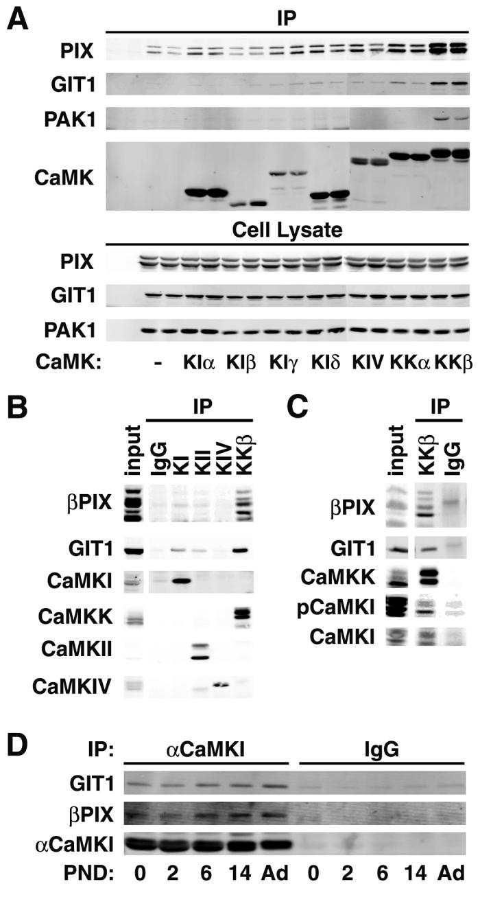
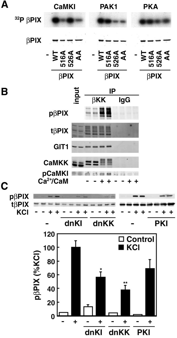
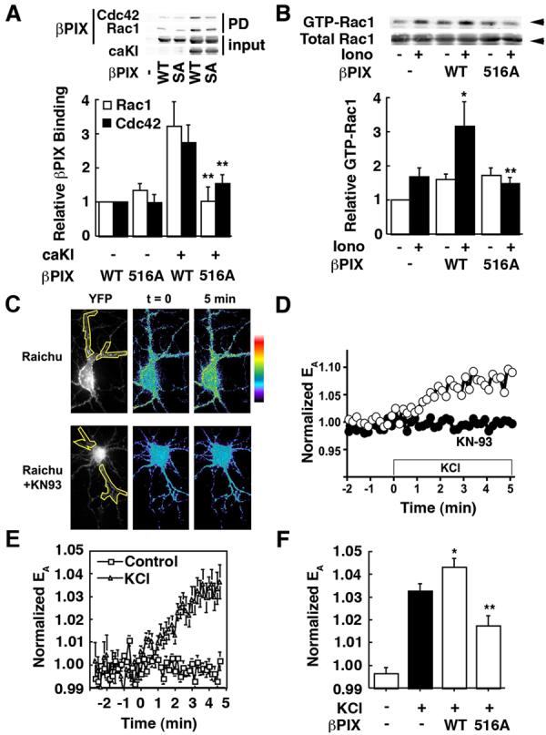
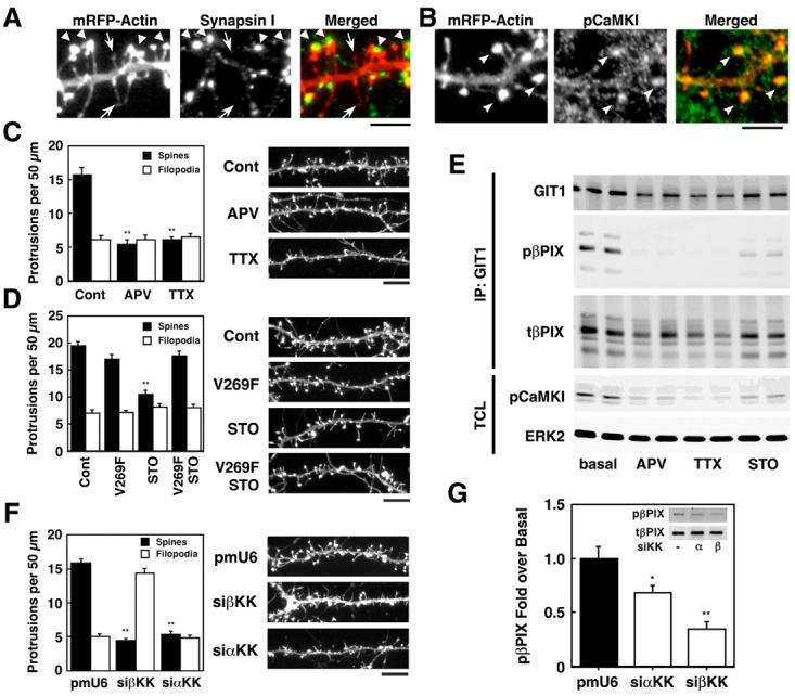
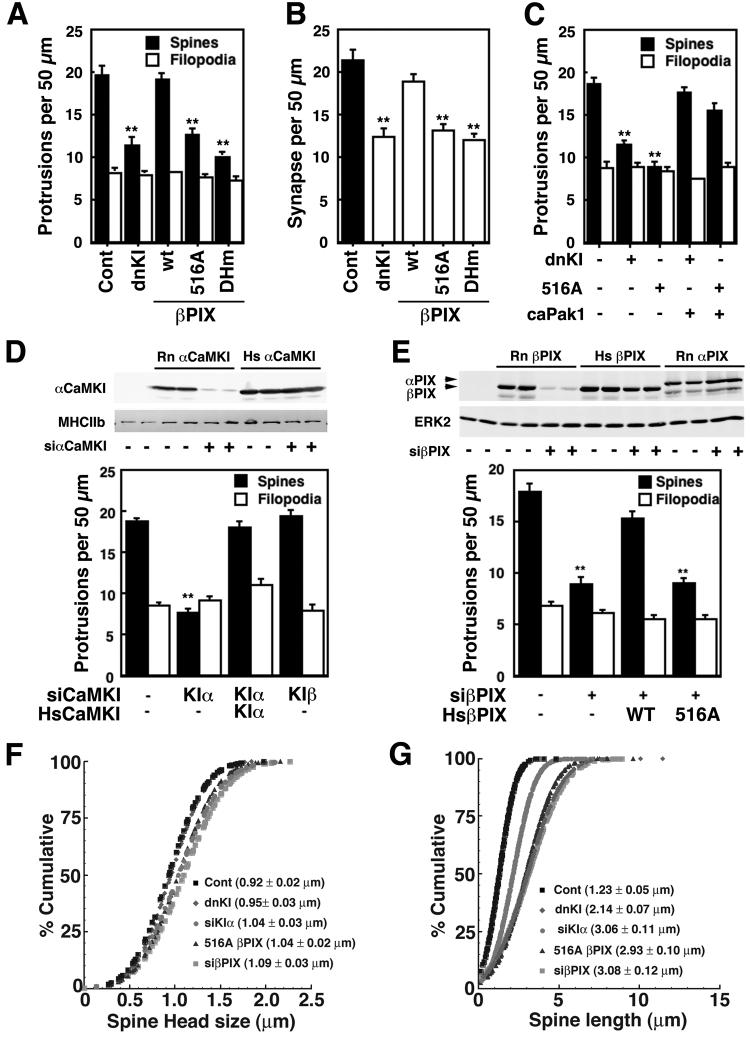
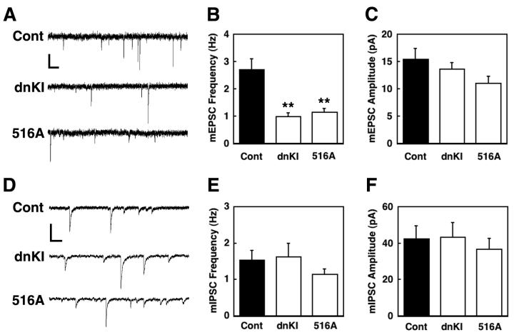
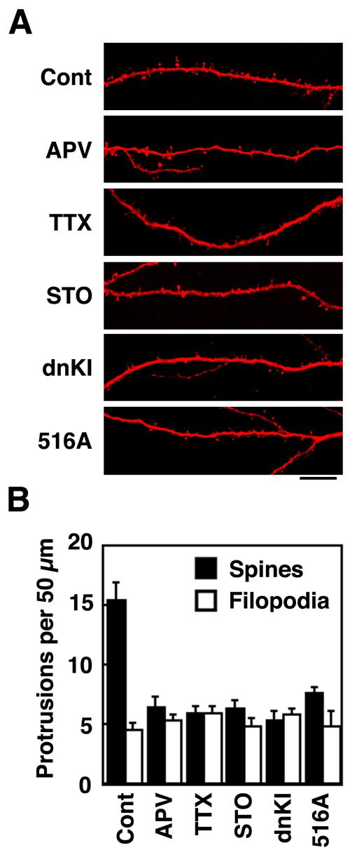
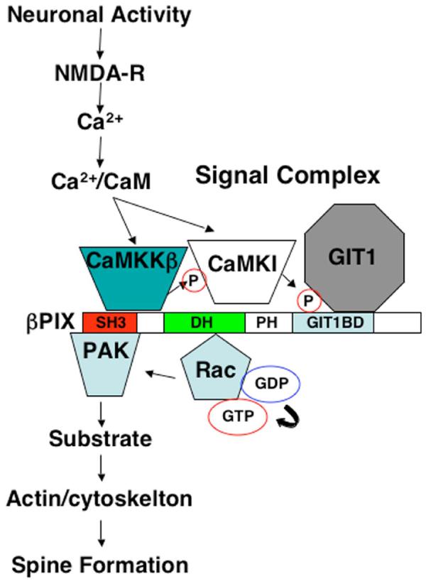
Neuronal activity augments maturation of mushroom-shaped spines to form excitatory synapses, thereby strengthening synaptic transmission. We have delineated a Ca(2+)-signaling pathway downstream of the NMDA receptor that stimulates calmodulin-dependent kinase kinase (CaMKK) and CaMKI to promote formation of spines and synapses in hippocampal neurons. CaMKK and CaMKI form a multiprotein signaling complex with the guanine nucleotide exchange factor (GEF) betaPIX and GIT1 that is localized in spines. CaMKI-mediated phosphorylation of Ser516 in betaPIX enhances its GEF activity, resulting in activation of Rac1, an established enhancer of spinogenesis. Suppression of CaMKK or CaMKI by pharmacological inhibitors, dominant-negative (dn) constructs and siRNAs, as well as expression of the betaPIX Ser516Ala mutant, decreases spine formation and mEPSC frequency. Constitutively-active Pak1, a downstream effector of Rac1, rescues spine inhibition by dnCaMKI or betaPIX S516A. This activity-dependent signaling pathway can promote synapse formation during neuronal development and in structural plasticity.


Figures

Figure 1
Figure 1
βCaMKK and αCaMKI interact with the βPIX/GIT1 complex. A. Co-immunoprecipitation from HEK293 cells. HEK293 cells were transfected with α (upper band) and βPIX (myc-tagged), GIT1 (EGFP fusion), Pak1 (Xpress-tagged) and the indicated FLAG-tagged CaMK. Duplicate cell lysates were immunoprecipated with FLAG antibody, washed, and western-blotted for the proteins indicated on the left. All of the transfected proteins were expressed to similar levels (see cell lysate) under all protocols. B. Co-immunoprecipitation from rat brain lysate. Brain lysates from P6 rat were immunoprecipitated with antibodies to αCaMKI, CaMKII, CaMKIV or βCaMKK, immunoprecipitates were washed and western-blotted for the proteins indicated on the left. C. Co-immunoprecipitation from rat hippocampus. Hippocampal lysates from P12 rat were immunoprecipitated with antibody to βCaMKK, washed and western-blotted for βPIX, GIT1, CaMKK, CaMKI, and phosphoCaMKI (pCaMKI). D. Co-precipitation of βPIX and GIT1 with αCaMKI. Brain lysate from the indicated postnatal days (PND) were immunoprecipitated with antibody to αCaMKI, washed and western blotted.
Figure 2
Figure 2
CaMKI phosphorylates Ser516 in βPIX. A. In vitro phosphorylation. Purified βPIX, wild-type (WT) or the indicated mutants (S516A, 516A; T526A, 526A; S516AT526A, AA), were subjected to in vitro phosphorylation using γ[32P]ATP and purified αCaMKI (activated by βCaMKK), PKA or Pak1. Reaction products were subjected to SDS/PAGE and autoradiography. B. Phosphorylation of βPIX Ser516 by CaMKI in the isolated multiprotein signaling complex. Brain homogenates from P6 rats were immunoprecipitated with anti-βCaMKK, washed, and incubated in the presence of Mg2+/ATP without or with Ca2+/CaM, subjected SDS/PAGE and western-blotted for phospho-βPIX, GIT1, CaMKK and phospho-CaMKI (pCaMKI). C. Phosphorylation in hippocampal neurons. Cultured hippocampal neurons (7DIV) were transfected with myc-βPIX and dnCaMKK, dnCaMKI or the PKA inhibitor protein PKI and treated with 0.5 μM tetrodotoxin in ACSF for 2 hrs to suppress neuronal activity. They were then incubated for 5 min with 56 mM KCl to activate Ca2+-dependent protein kinases and PKA. Cell lysates were immunoprecipitated with myc-antibody and western-blotted for phospho-S516 βPIX. *P<0.05, **P<0.001 compared to control.
Figure 3
Figure 3
CaMKI-dependent phosphorylation of S516 stimulates βPIX GEF activity. A. Interaction of Rac/Cdc42 with βPIX. HEK293 cells were transfected with myc-βPIX (WT or S516A) and caCaMKI (caKI) as indicated. Cell lysates were mixed with GST-Rac1T17N or GST-Cdc42NT17N and the GST complex isolated by glutathionine-Sepharose. The myc-βPIX that co-purified with the GST-Rac1 or GST-Cdc42 was quantified by western blot. B. GEF activity of βPIX in HEK293 cells. HEK293 cells were transfected with βPIX (WT or S516A) and myc-Rac1 and treated for 3 min without or with 1 μM ionomycin. Cell lysates were mixed with GST fused to the CRIB domain of Pak1 (residues 67-150) which specifically binds to activated (i.e., GTP-loaded) Rac, and the GST complex was isolated by glutathione-Sepharose (Soderling et al., 2002). Bound Rac1 was quatified by western blotting. *P< 0.05 compared to control, **P< 0.05 compared to wild-type. C-E. Depolarization increases CaMK-dependent Rac GEF activity in hippocampal neurons. Hippocampal neurons, transfected with Raichu-Rac1, were live-imaged upon stimulation by 28 mM KCl without or with 1 hr pretreatment with the general CaMK inhibitor KN-93 (5 μM, panel C lower images, panel D solid circles). Normalized FRET efficiencies (EA, see Methods) or YFP signal (panel C, far left; region of proximal dendrites analyzed are outlined in yellow) are shown for representative cells (C, D) or multiple cells (E, n=15). See Fig. S4 for an enlargement of the horizonal dendrite in top of panel C. F. GEF activity of transfected βPIX in neurons. mRFP-βPIX WT (WT) or S516A mutant (516A) were co-transfected with Raichu-Rac1 (ratio 5:1) into hippocampal neurons. FRET efficiencies (EA) in proximal dendrites were averaged from 5 time points between 4-5 min. *P< 0.05 compared to Raichu alone, **P< 0.001 compared to wild-type (n=6-17).
Figure 4
Figure 4
CaMKK modulates activity-dependent dendritic spine density and phosphorylation of βPIX. A, B. Cultured hippocampal neurons (7 DIV) were transfected with mRFP-βactin to visualize dendritic protrusions, fixed on DIV 14, and immunostained for the presynaptic marker synapsin 1 (A) or pCaMKI (B). A. The left panel shows mRFP-βactin; middle panel, synapsin 1; right panel, overlay. Spines (large arrows) were identified as mushroom-shaped projections with intense expression of mRFP-βactin at their tips (heads) whereas filopodia (small arrows) were thin with low expression of mRFP-βactin throughout. (Scale bar, 5 μm). B. The left panel shows mRFP-βactin; middle panel, phosphoCaMKI (pCaMKI); right panel, overlay. PhosphoCaMKI signals were identified in spines (arrow heads) (Scale bar, 5 μm). C,D,F. DIV 7 neurons were transfected with mRFP-βactin without or with the STO-insensitive mutant of βCaMKK (V269F) (D) or siRNAs specific for α or βCaMKK (F). Where indicated, neurons were treated for 4 days with 100 μM APV (block NMDAR), 1 μM TTX (inhibit neuronal activity) or 20 μM STO-609 (inhibit CaMKK). On 12 DIV (C, D) or 14 DIV (F) the neurons were fixed, dendrites were imaged and at least three different 50 μm sections per neuron (15-20 neurons per condition) were analyzed for protrusions. Representative dendrites are shown on the left, and summaries of the effects of transfections or pharmacological treatments on protrusion densities are shown on the right. **P<0.001, compared to control. Scale bar, 10 μm. E. On 12DIV, the neurons were lysed, immunoprecipitated with GIT1 antibody, western-blotted with GIT1, phospho-βPIX (pβPIX), βPIX (tβPIX), phosphoCaMKI (pCaMKI), and ERK2 (as loading control). G. DIV 7 neurons were transfected with myc-βPIX with or without siRNAs specific for α or β CaMKK. On 11 DIV, neurons were lysed, immunoprecipitated with myc antibody, western-blotted with phospho-βPIX (pβPIX) and myc (tβPIX). *P<0.05, **P<0.001 compared to control.
Figure 5
Figure 5
Role of CaMKI and βPIX S516 phosphorylation on spinogenesis. A-C. Cultured hippocampal neurons (DIV 7) were transfected with mRFP-βactin plus either dnCaMKI or βPIX (WT, S516A or a mutant (DHm) devoid of GEF activity) ± caP ak1 as indicated. On DIV 14 neurons were fixed, stained for synapsin 1 and images were analyzed for density of spines and filopodia (A). Synapses (B) were identified as spines with adjacent staining of synapsin 1 (see Fig. S10). D, E. Neurons (DIV 7) were transfected with mRFP-βactin plus siRNAs for rat (Rn) α or βCaMKI (D) or βPIX (E) ± plasmids expressing human αCaMKI or human (Hs) βPIX (WT or S516A) as indicated. Spines/filopodia were analyzed as in Fig. 4 (**P<0.001, compared to control). HEK293 cells were transfected with Flag-tagged αCaMKI from rat (Rn) or human (Hs) plus siRNA for rat αCaMKI, lysed and immunoblotted for Flag and myosin heavy chain IIb (MHCIIb, loading control) (D, top panel). HEK293 cells transfected with Myc-tagged βPIX from rat or human or Myc-tagged rat αPIX plus siRNA for rat βPIX were immnoblotted for Myc and ERK2 (loading control), as indicated (E, top panel). F, G. Neurons (DIV 7) were co-transfected with mRFP-βactin plus other constructs as indicated, fixed on DIV14 and analyzed for spine head width (F) and length (G).
Figure 6
Figure 6
Effect of dnCaMKI and βPIX S516A mutant on mEPSCs and mIPSCs. Cultured neurons (DIV 7) were transfected with EYFP-γactin and either mRFP (controls), mRFP-dnCaMKI or mRFP-βPIX S516A mutant, and mEPSCs and mIPSCs were recorded on DIV 12. Representative traces of mEPSCs (A) or mIPSCs (D) recorded from control (Cont), dnCaMKI-expressing neuron (dnKI), or βPIX S516A-expressing neuron (516A). Frequencies (B, E) and amplitudes (C, F) of mEPSCs or mEPSCs. **P<0.005, compared to control. Note that suppression by dnCaMKI and βPIX S516A of mEPSCs, but not mIPSCs, correlates with their effects on synapse number from Fig. 5B. Scale bars: top, 20 pA and 250 ms; bottom, 100 pA and 250 ms.
Figure 7
Figure 7
Regulation of spinogenesis in cultured hippocampal slices Hippocampal slices from P7 rats were cultured for 2 days, subjected to biolistic transfection with mRFP-actin ± other plasmids as indicated. Neurons were treated without or with pharmacological reagents (100 μM APV, 1 μM TTX, or 20 μM ST O-609) as indicated. On DIV 5, dendritic protrusions were analyzed for spines and filopodia as in Fig. 5. A. Representative examples of apical CA1 dendrites from pyramidal neurons in hipocampal organotypic slice culture. Scale bar, 15 μm. B. Summary of the effects of transfections and pharmacological treatments on protrusion density.
Figure 8
Figure 8
Model of CaMK/βPIX signaling pathway. Activity-dependent Ca2+ influx through the NMDA receptor of a spine activates the CaMKK/CaMKI/βPIX complex to promote CaMKI-mediated phosphorylation of S516 in βPIX with resultant activation of its GEF activity. The activated βPIX promotes GTP-loading of Rac (and/or Cdc42) to activate Pak and modulate actin dynamics and enhance spinogenesis and synapse formation.

Comment in

Similar articles

Cited by

References

    1. Ackermann M, Matus A. Activity-induced targeting of profilin and stabilization of dendritic spine morphology. Nat Neurosci. 2003;6:1194–1200. - PubMed
    1. Audebert S, Navarro C, Nourry C, Chasserot-Golaz S, Lecine P, Bellaiche Y, Dupont JL, Premont RT, Sempere C, Strub JM, et al. Mammalian Scribble forms a tight complex with the betaPIX exchange factor. Curr Biol. 2004;14:987–995. - PubMed
    1. Bahr BA. Long-term hippocampal slices: a model system for investigating synaptic mechanisms and pathologic processes. J Neurosci Res. 1995;42:294–305. - PubMed
    1. Bokoch GM. Biology of the p21-activated kinases. Annu Rev Biochem. 2003;72:743–781. - PubMed
    1. Bonhoeffer T, Yuste R. Spine motility. Phenomenology, mechanisms, and function. Neuron. 2002;35:1019–1027. - PubMed

Publication types

MeSH terms

Substances